- 1IAS, CNRS, Orsay, France (rosario.brunetto@ias.u-psud.fr)
- 2DIST-Università Parthenope, Napoli, Italy
- 3INAF-IAPS, Roma, Italy
- 4SOLEIL synchrotron, Gif-sur-Yvette, France
- 5Division of Earth and Planetary Materials Science, Graduate School of Science, Tohoku University, Japan
- 6Institute of Space and Astronautical Science (ISAS), Japan Aerospace Exploration Agency (JAXA), Sagamihara, Japan
- 7Institut d’Electronique, de Microélectronique et de Nanotechnologie, Lille, France
- 8Research Organization of Science and Technology, Ritsumeikan University, Kyoto, Japan
- 9Guangzhou Institute of Geochemistry, Guangdong, China
1. Introduction
The Hayabusa2/JAXA spacecraft has orbited and studied near-Earth object Ryugu [1]. Two sample collections have been performed and the spacecraft is currently on its way back to the Earth for an expected return in late 2020. A preliminary examination phase will follow [2], expected to elucidate the formation and evolution of Ryugu. Considering the limited amount of material that will be retrieved, a multi-analytical sequence is needed to maximize the scientific outcome and minimize sample loss. Among the possible laboratory techniques, IR spectroscopy is important in being totally non-destructive and comparable to remote sensing observations of small bodies [3]. Thanks to IR imaging micro-spectroscopy, it is possible to detect and study the spatial distribution of molecular bonds associated to minerals, water and organic compounds, and their co-localization [4]. We consider IR three dimensional (3D) micro-tomography (IR-CT) an excellent starting point in a multi-analytical sequence to be applied on returned samples [5]. Here we report IR hyperspectral measurements of carbonaceous chondrites as a rehearsal of IR-CT and IR hyperspectral imaging that will be part of the multi-analytical sequence of the “MIN-PET CG” Hayabusa2 team (mineralogy and petrology of coarse grains) led by T. Nakamura.
2. Methods
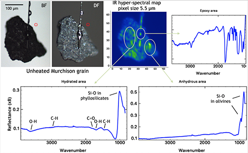
We performed IR measurements in reflectance on bulk fragments of selected meteorites, and both in reflectance and transmittance on several isolated grains (sizing 20-50 µm) extracted from carbonaceous chondrites. We also analyzed individual grains of the Murchison CM meteorite prepared at Tohoku University (Japan) from three bulk samples: (1) unheated, (2) heated at 400°C, and (3) heated at 600°C [6]. The laboratory-controlled heating was applied to simulate potential heating undergone by Ryugu surface materials, as suggested by some Hayabusa2 observations [7]. Some of these Murchison grains were mounted on tungsten needles by means of a platinum welding performed in IEMN-Lille (France) with a focused ion beam microscope. Other grains were prepared at Tohoku University, mounted on carbon fibers using epoxy (Fig. 1).
We analyzed the samples using an IR hyperspectral imaging and micro-tomography setup installed at the SMIS beamline of the SOLEIL synchrotron (France). This setup has already been used for analyzing Hayabusa samples from asteroid Itokawa [8]. FTIR data were collected using an Agilent Cary 670/620 microspectrometer. In transmission mode we used a X25 objective coupled with high magnification optics (providing an additional X2.5 magnification) placed in front of a 128x128 pixels FPA detector, to obtain a projected pixel size of ~0.66 µm on the focal plane, and a field of view of ~84 µm. IR-CT is performed in transmission mode using the method described by Dionnet et al. [8]. In reflection mode we used a X15 objective, with a projected pixel size of ~5.5 µm on the focal plane, and a field of view of ~700 µm (Fig. 1). In both cases we collected hyperspectral data: for each pixel we obtained an IR spectrum in the 850-3950 cm-1 spectral range. The spatial resolution was diffractionlimited for the whole investigated spectral range.
3. Results
Infrared spectra show the presence of bending and stretching absorption bands of chemical bonds (C-H, OH, Si-O, C=O, etc.) of different functional groups, as expected from literature IR spectra of Murchison and other chondrites [9,10]. The relative intensities of these bands are found to vary among different grains, and their 3D spatial distribution is heterogeneous within individual grains (see Fig. 1). Noticeable differences are found between the IR spectra of unheated and heated Murchison samples, with a general trend of increasing the anhydrous to hydrated silicate content with increasing temperature, and reducing the organic content.

Fig. 1. A typical IR reflectance hyperspectral map of an unheated Murchison sample mounted on a carbon needle (top left, microscope images in bright and dark field), with the detection of hydrated (bottom left) and anhydrous (bottom right) areas. The epoxy contribution can be clearly separated (top right).
4. Summary and Conclusions
IR data provide a first quick look at the composition, abundance and 3D distribution of mineral phases and carbonaceous materials at the scale of a few micrometers. Once regions of interest are identified by IR measurements, thin sliced sections of the samples can be analyzed by more destructive techniques to retrieve the structure and the elemental and isotopic composition of the carbonaceous component and its mineral host, down to the nanometer scale [11]. In addition, the IR data are useful in the comparison with remote sensing observations of asteroid surfaces [7,12]. This top-down sequence will help us building a bridge between the remote sensing and in situ observations of Ryugu at macroscopic scale and the chemical and physical processes operating at the nanoscale.
Acknowledgments
The micro-spectroscopy measurements were supported by grants from Region Ile-de-France (DIM-ACAV) and SOLEIL. This work has been funded by the CNES (France) and by the ANR project CLASSY (Grant ANR-17-CE31-0004-02) of the French Agence Nationale de la Recherche. This work was partly supported by the French RENATECH network.
References
[1] Watanabe S. et al. (2019) Science 364, 268-272. [2] Tachibana S. et al. (2018) AGU Fall Meeting, abstract #P33C-3846. [3] Brunetto R. et al. (2011) Icarus 212, 896–910. [4] Dionnet Z. et al. (2018) Meteoritics & Planet. Sci. 53, 2608-2623. [5] Dionnet Z. et al. (2018) Microscopy and Microanalysis 24, 2100-2101. [6] Mogi K. et al. (2017) 80th Annual Meeting of the Meteoritical Society, Abstract #6225. [7] Kitazato K. et al. (2019) Science 364, 272-275. [8] Dionnet Z. et al. (2020) Meteoritics & Planet. Sci., in press. [9] Lantz C. et al. (2015) A&A 577, A41. [10] Beck P. et al. (2014) Icarus 229, 263-277. [11] Aléon-Toppani A. et al. (2020) Lunar and Planetary Science Conference 2682. [12] Hamilton V. et al. (2019) Nat. Astron. 3, 332–340.
How to cite: Brunetto, R., Aléon-Toppani, A., Dionnet, Z., Rubino, S., Arribard, Y., Baklouti, D., Borondics, F., Djouadi, Z., Lantz, C., Matsumoto, M., Matsuoka, M., Nakamura, T., Amano, K., Takahashi, M., Troadec, D., and Tsuchiyama, A.: Hyperspectral imaging of carbonaceous chondrites in view of the Hayabusa2 sample return, Europlanet Science Congress 2020, online, 21 Sep–9 Oct 2020, EPSC2020-121, https://doi.org/10.5194/epsc2020-121, 2020.

