
 1,2,1,3,1,6,5,
- 1Centre for X-ray Tomography (UGCT), Ghent University, Gent, Belgium (matthieu.boone@ugent.be)
- 2Dept. Physics and Astronomy, Ghent University, Gent, Belgium
- 3Dept. Geology, Ghent University, Gent, Belgium
- 4Dept. Chemistry, Ghent University, Gent, Belgium
- 5European Space Agency (ESA), Mars Science Team (HRE-MS), Noorderwijk, The Netherlands
- 6Max Planck Institute for Solar System Research, Göttingen, Germany
1. Introduction
The NASA/ESA Mars Sample Return (MSR) Campaign aims to bring Martian rock and soil specimens to Earth for high-priority scientific investigations, marking a landmark achievement in planetary exploration [1]. As part of the MSR Sample Receiving Project (SRP) Pre-Basic Characterization Phase, these samples will first undergo non-destructive analysis inside a dedicated Sample Receiving Facility (SRF) while still enclosed in precision-engineered titanium tubes. One of the first techniques applied will be high-resolution X-ray computed tomography (HR XCT), used to non-destructively generate detailed 3D digital representations of samples. These digital twins will contribute to a comprehensive sample catalogue, supporting subsample allocation through competitive, open science announcements. To ensure XCT delivers maximal structural information at micron-level resolution while preserving sample integrity, it is critical to optimize scanning protocols—balancing image quality with minimal radiation dose. This initiative lays the groundwork for curatorial excellence and scientific readiness, ensuring that returned Martian materials are studied using state-of-the-art methods while preserving their pristine condition for future research. This ongoing study describes the first imagery, data simulation and dose simulation results, alongside the feasibility of scanning within the Sample Tube Isolation Container (STIC).
2. Methodology & Results:
XCT Imaging
Test scans of two types of analogue test samples (i.e., loose unconsolidated rocks and solid rock cores) were performed using the High-Energy CT system Optimized for Research or HECTOR [2] at the Ghent University Centre for X-ray Tomography (UGCT). Starting with a low-resolution scan (60 µm) of the complete tube and a high-resolution scan (7µm) of the tube filled with loose unconsolidated rocks, a resolution of 10 µm was chosen as voxel size for the other datasets. The voltage (120 kV) was set to match bench-top sized HR XCT systems to be potentially used in the SRF. The same settings have been applied to image the solid rock cores in their sample tubes as well as within the STIC.
Simulations of datasets
Simulations of virtual samples have been recreated by segmenting the loose grain and solid rock from the surrounding air and tubing from HR XCT images of the analogue test samples. Using the in-house developed realistic projection simulator [3], mineral phases have been recreated to anticipate all possible mineral compositions of the Mars samples [4]. The HR XCT images of these virtual samples have been used to assess the image quality (contrast) and application potential in predicting the composition of the unopened samples.
Dose simulations
To simulate the dose deposition in the samples during XCT scanning, a workflow was developed using the Monte Carlo code PENELOPE [5]. The cone-beam geometry as well as the polychromatic X-ray beam is taken into account.
3. Discussion:
The sample tubes of well-defined dimensions largely determine the boundary conditions under which the samples can be optimised. The diameter of the sample tube, and the STICs around the tube, set the limitations on the achievable voxel size to be around 10 µm. On the other hand, the titanium material of the sample tube serves as an excellent filter and omits the need for extra hardware beam filtering, although such filter may reduce the radiation dose. Visible image artefacts seem to be rather caused by the irregular shape of the tube (especially the fin at the bottom of the tube) but remain appreciably low for the investigation of the rock material within.
The contrast between the different mineral phases is improved by lowering the initial voltage of the X-ray source. A voltage of 120 kV is considered to be a good trade-off between high image quality and low image artefacts due to beam hardening. The projection simulator enabled the prediction of the relative gray values of the mineral phases and can be used to better predict the mineral content of the unopened samples. Optimizing the differentiation between specific minerals of interest could benefit from further adjusting the HR XCT settings or by using hyperspectral HR XCT.
4. Conclusions:
From the preliminary results, a resolution of 10µm is sufficient to retrieve valuable information from the Martian samples in the SRF, prior to opening the tubes. However, careful assessment of the deposited dose and the effects thereof is still needed.
Acknowledgements
This study is funded by ESA under ESA contract number AO/1-12041/23/NL/AT.
References:
[1] G. Kminek et al., 2022; [2] Masschaele et al., 2013; [3] Dhaene et al., 2015; [4] Morrison et al. 2024; [5] Baró et al., 1995
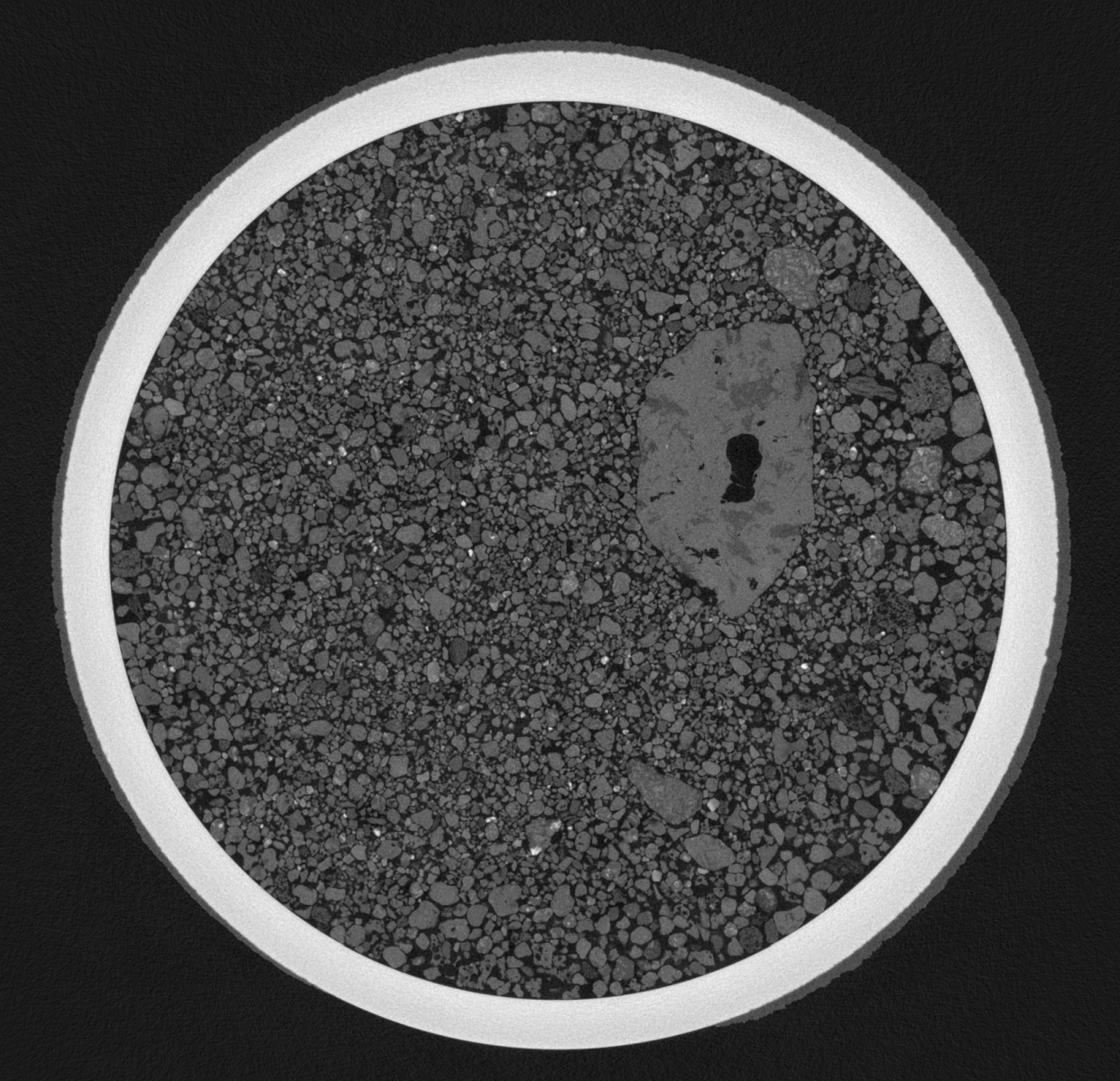
Example micro-CT slice of the sample tube with a granular analogue sample, scanned at Ghent University (voxel size: 10 µm)
How to cite: Boone, M. N., Buyse, F., Baert, T., Vincze, L., Josipovic, I., Tack, P., Van Hoorebeke, L., Zemeny, A., Thiessen, F., Sefton-Nash, E., and Kminek, G.: Pre-basic characterizations for the Mars Sample Return Sample Receiving Project: preliminary results of X-ray micro-CT analysis, EPSC-DPS Joint Meeting 2025, Helsinki, Finland, 7–13 Sep 2025, EPSC-DPS2025-1504, https://doi.org/10.5194/epsc-dps2025-1504, 2025.