Multiple terms: term1 term2
red apples
returns results with all terms like:
Fructose levels in red and green apples
Precise match in quotes: "term1 term2"
"red apples"
returns results matching exactly like:
Anthocyanin biosynthesis in red apples
Exclude a term with -: term1 -term2
apples -red
returns results containing apples but not red:
Malic acid in green apples
hits for "" in
Network problems
Server timeout
Invalid search term
Too many requests
Empty search term
MITM13
Laboratory work on cosmo-materials either fallen on earth, returned by space missions or synthetically produced are essential for the understanding of the history of planets, moons, and small bodies. The results of controlled experiments are essential for the interpretations of measurements obtained by ground-based and space observations as well as exploration missions. They are also necessary for planning and preparing future in situ and sample-return space missions, ensuring their success in collecting valuable samples and data.
In this session, we invite submissions related to the analysis of cosmo-materials and to the production, evolution and analysis of planetary, moons and small bodies analogues (interpretation of chemical/physical properties, predictions, preparation of analytical tools or space instruments, preparation of analytical chain for sample return analyses, etc.). Laboratory experiments necessary to interpret data of any past, present and future space missions will be particularly encouraged.
A primary objective of the NASA Mars 2020 Perseverance rover mission is the detection of organic matter on Mars, which may provide key insights into the past habitability of Mars and the potential presence of past life. Indeed, molecular biosignatures‒organic molecules diagnostic of biological processes‒may provide more direct evidence of biogenicity than other categories of biosignatures for which biological production is only inferred. However, the degradation of the molecules in the harsh Martian environment over time, along with the limitations of the space flight instruments and the possible transformations induced by the measurements themselves, makes it quite challenging to identify the original material and differentiate between biotic and abiotic organic matter.
The Perseverance rover utilizes a specialized suite of instruments to map minerals and organic distributions at a microscopic scale. Notably, the Scanning Habitable Environments with Raman and Luminescence for Organics and Chemicals (SHERLOC) instrument, a deep UV (DUV) Raman and fluorescence spectrometer, previously identified Raman features in the Quartier abrasion target on the Jezero crater floor that suggest the presence of aromatic organics within sulfate matrices [1].
This study builds upon previous findings by reporting similar Raman features in the Pilot Mountain target, located on the Jezero fan top. To interpret these signals, we carried out a systematic laboratory investigation and compared these SHERLOC observations with laboratory data from reference organo-sulfate samples to evaluate the possible organic sources for the detected spectroscopic features [2]. Specifically, we prepared organo-sulfate analog samples using a variety of aromatic organic compounds easily detectable by SHERLOC (both abiotic and of potential biotic origin), and characterized them with laboratory DUV Raman and fluorescence spectrometers analogous to SHERLOC, i.e. the ACRONM instrument located at NASA’s Johnson Space Center, the Brassboard instrument located at the Jet Propulsion Laboratory, and the Photon Systems RPL200 instrument located at INAF-Astrophysical Observatory of Arcetri, operated to mimic SHERLOC analysis.
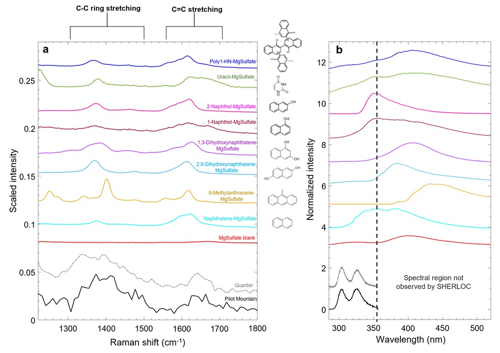
Comparison between our laboratory dataset and SHERLOC observations for Quartier and Pilot Mountain shows that different types of aromatic organics in sulfates tested in this work‒naphthalene, 1- and 2-naphthol (HN), 1,3- and 2,6-dihydroxynaphthalene (DHN), 9-methylanthracene (9-MA), uracil and an oxy-PAH polymer synthesized from 1-HN (poly1-HN)‒exhibit intense C-C and C=C ring stretching bands (and C=O stretching bands in the case of compounds like uracil with carbonyl functional groups on the aromatic ring) at a variety of positions within the spectral regions of interest, though none of the tested organo-sulfate analogs exhibit fluorescence resembling the co-located fluorescence doublet at ~303 and 325 nm observed in Quartier and Pilot Mountain (Fig. 1). However, the polycyclic aromatic hydrocarbons (PAHs) 9-MA, 2,6-DHN and 1,3-DHN in magnesium sulfate fluoresce mostly outside the SHERLOC region, which may be consistent with an inorganic origin of the co-located fluorescence doublet, such as luminescence of Ce3+ in anhydrite [1,3], illustrating that similar types of three/two-ring aromatic organic molecules could be present.

Fig. 1. Cross-comparison of the features of interest in Quartier and Pilot Mountain with spectra of organo-sulfate analogs for Raman (a) and fluorescence (b).
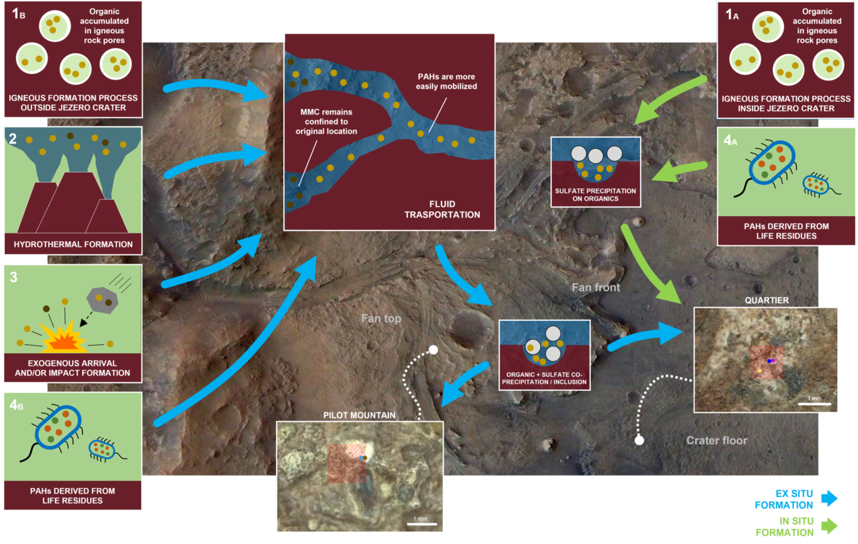
The association with igneous lithologies suggests that PAHs formed endogenously by igneous processes (Fig. 2). Their location in pores of the igneous rocks may be consistent with Fischer-Tropsch-like synthesis from trapped magmatic gases catalyzed by iron oxides. PAHs might have been subsequently preserved by sulfates precipitated in the pores of the igneous rocks as consequence of their aqueous alteration. Such salt-mediated preservation is particularly efficient on Earth, since sulfate minerals can trap organics within intracrystalline inclusions [4], and exhibit photoprotective properties [5,6].
Regional hydrothermal groundwaters associated with Syrtis Major volcanism might also have played a role in sulfate formation and PAH transportation, as well as in dissolving igneous rocks causing mobilization of Ce3+ and its transportation through fractures and precipitation as sulfate veins, which would explain the co-presence of Ce3+ and PAHs in these sulfates (Fig. 2).
The organo-sulfate association is consistent with studies on Martian meteorites [7] and observations from Gale crater [8] and reinforces the hypothesis that sulfates might have been key in the preservation and transport of organic molecules in the Martian environment and, hence, could have played a significant role in the Martian carbon cycle, influencing the availability and cycling of carbon compounds necessary for life.
Although we have not found evidence of biogenicity, we cannot exclude that the organics detected in these rocks might be resistant alteration products from the chemical weathering of ancient biotic compounds, and/or a correlation with the organic carbon of potential biotic origin detected in the Bright Angel formation [9]. With the limitations of Perseverance’s Raman and fluorescence spectroscopy techniques, addressing these various hypotheses would require further laboratory studies to retrace the chemical weathering processes that might have occurred at Jezero crater over time leading to the formation of the observed organics, and returning these samples to Earth for high sensitive analysis in Earth-based laboratories.

Fig. 2. Potential mechanisms for the presence of PAHs in sulfates in Quartier and Pilot Mountain.
Acknowledgments: This research was supported by the Italian Space Agency (ASI) through the ASI/INAF agreement no. 2025-12-HH.0, and by INAF Large Grant 2024.
References: [1] Sharma S. et al. (2023) Nature, 619, 724–732. [2] Fornaro T. et al. (2025) Nature Astronomy, 9, 1648–1661. [3] Haney N. C. et al. (2026) JGR-Planets, 131(3), e2025JE009385. [4] Sephton M. A. et al. (2023) J Geol Soc London, 180. [5] Alberini, A. et al. (2024) Scientific Reports, 14, 15945. [6] Alberini A. et al. (2025) Scientific Reports, 15(1), 40484. [7] Jaramillo E. A. et al. (2019) Geophys Res Lett, 46, 3090–3098. [8] Eigenbrode J. L. et al. (2018) Science, 360, 1096–1101. [9] Hurowitz, J. A. et al. (2025) Nature, 645(8080), 332–340.
How to cite: Fornaro, T. and the Organo-Sulfate Analog Study Working Group: Aromatic Organic Compounds Associated with Sulfates at Jezero Crater: Insights from SHERLOC and Laboratory Analogs, Europlanet Science Congress 2026, The Hague, The Netherlands, 7–11 Sep 2026, EPSC2026-194, https://doi.org/10.5194/epsc2026-194, 2026.
Introduction
Mars is one of the primary targets of astrobiological exploration, due to the presence of liquid water on its surface during its early stages and the existence of sedimentary environments that may have hosted and preserved biosignatures [1]. One of the main aims of the current and future missions, including NASA Mars 2020 Perseverance and ESA ExoMars Rosalind Franklin rovers, is to detect such biosignatures using advanced spectroscopic instruments operating across multiple spatial scales [2][3]. To fully exploit these datasets, it is essential to establish a robust interpretive framework based on the characterization of analogous terrestrial samples and Martian meteorites. Within this framework, terrestrial analog environments, such as Proterozoic clay-rich mudstones from the ~1 Ga El Mreiti Group (Mauritania), allow for the study of well-preserved biosignatures formed under sedimentary and diagenetic conditions comparable to those hypothesized for early Mars [4]. At the same time, Martian meteorites offer direct evidence of Martian geochemical evolution and the alteration of organic matter, albeit modified by secondary processes such as thermal and shock alteration during ejection from Mars and entry in Earth Atmosphere [5]. The combined study of terrestrial analogs and Martian meteorites is therefore essential for defining the pathways of biosignature preservation and assessing their detectability in Martian contexts. Another key aspect is the use of techniques that closely mimic those employed in current and future Mars missions. By applying spectroscopic methods relevant to these missions to complex natural samples, it becomes possible to assess detection limits, spectral ambiguities, and the influence of mineral matrices on the expression of biosignatures, thereby providing direct support for the interpretation of in situ data.
Methods
A multi-scale spectroscopic approach was implemented using techniques analog to those deployed on Mars 2020 and ExoMars rovers. Bulk mineralogical and compositional information was obtained through Fourier Transform Infrared (FTIR) spectroscopy in reflectance mode, enabling the identification of dominant mineral phases and broad organic functional groups. Micro-scale heterogeneity was investigated using a Hyperion 1000 micro-FTIR system, allowing the spatial mapping of mineral–organic associations. Visible and Deep Ultraviolet (DUV) Raman spectroscopy and fluorescence analyses were employed to probe the molecular structure and distribution of organic compounds, in a mineral matrix, with particular sensitivity to aromatic and conjugated systems such as kerogens and macromolecular carbons (MMC). This integrated methodology enables a systematic evaluation of biosignature detectability across analog and extraterrestrial materials.
Results
The results presented in this study provide a framework for understanding the spectroscopic expression of biosignatures in complex natural materials. Indeed, the investigation of terrestrial analog samples and Martian meteorites yields key insights into how biosignatures may be preserved, modified, and detected under realistic geological conditions. In particular, this work highlights the importance of multi-scale characterization in resolving mineral–organic associations, demonstrating how biosignatures may manifest differently depending on spatial resolution and analytical technique. The datasets contribute to defining practical detection limits for mission-relevant instruments, suggesting which spectroscopic approaches are best suited for identifying specific classes of organic compounds and their mineralogical context. Furthermore, the results provide constraints on the expected spectral signatures of biosignatures in Martian environments, offering guidance on how such signals may appear – or be obscured – within natural, heterogeneous matrices. Overall, this study establishes a foundation for both current and future analyses of Martian datasets, contributing to the development of more robust biosignature detection strategies and improving the reliability of interpretations derived from in situ and returned sample investigations.
References: [1] Vago J.L. et al. (2017), Astrobiology 17, 471–510; [2] Farley K.A. et al. (2020), Space Sci Rev 216, 142; [3] Vago J. et al. (2015), Solar System Research 49, 518–528; [4] Beghin J. et al. (2017), Precambrian Research 299, 1–14; [5] Steele A. et al. (2016), Meteorit Planet Sci 51, 2203–2225.
Acknowledgements: This research is supported by the Italian Space Agency (ASI) through the ASI/INAF agreement no. 2025-12-HH.0 and PAMMAT project “Alteration processes in Mars and Moon meteorites, and Terrestrial Analogues at different environments: Mars2020, Rosalind Franklin and Returned Samples from Mars and Moon” (Grant No. PID2022-142750OB-I00), funded by the Spanish Agency for Research MCIN/AEI/10.13039/501100011033/FEDER-UE.
How to cite: Alberini, A., Fornaro, T., García Florentino, C., Poggiali, G., Biancalani, S., Renzi, F., Coloma, L., Battistuzzi, M., Roussel, A., Aramendia, J., Madariaga, J. M., and Brucato, J. R.: Supporting the Search for Organics on Mars Through Mission-Analog Spectroscopic Characterization of Terrestrial Analogs and Martian Meteorites, Europlanet Science Congress 2026, The Hague, The Netherlands, 7–11 Sep 2026, EPSC2026-905, https://doi.org/10.5194/epsc2026-905, 2026.
Future Mars Sample Return missions will provide extremely limited quantities of rock and soil, posing challenges for conventional geotechnical testing, which typically requires larger specimen masses (e.g., 100s to 1000s of grams). Depending on the return material type, a single sample may contain only ~10 g of loose or regolith-like material. This study investigates how reduced specimen mass affects the results of common geotechnical index tests using the Mars regolith simulant MGS-1, focusing on water content, particle density, particle-size distribution (PSD), and loose/tapped bulk density. Reference values were first established using standard specimen masses (typically ~100 g or ASTM-recommended quantities). Tests were then repeated while systematically reducing the mass (10 g, 2 g, and 0.5 g) to evaluate sensitivity to specimen size. Results demonstrate that sensitivity to reduced mass is strongly method-dependent. At 10 g, most tests (water content, particle density, wet-prepared sieving, and bulk density) remained consistent with reference values and showed low variability. At 2 g, deviations and uncertainty increased, although some methods still followed expected trends. At 0.5 g, results were largely dominated by handling effects and measurement limitations. Dry sieving underestimated fines content compared to wet-prepared sieving and is not recommended as a stand-alone method. Hydrometer testing was identified as the main limitation for full PSD characterization, requiring larger masses for reliable results.
Overall, 10 g represents the lowest broadly reliable specimen mass for MGS-1 across most index tests, with limitations for fine-fraction PSD. While smaller masses may still yield useful data, confidence becomes increasingly method-dependent. The study demonstrates that meaningful geotechnical characterization is feasible with Mars Sample Return-scale materials, provided that deviations from standard methods and associated uncertainties are clearly documented.
How to cite: Setvik, S. H., Quinteros, S., Kim, T., Skurtveit, E., Mikesell, T. D., and Griffiths, L.: Specimen-mass sensitivity in geotechnical index testing of expexted Martian samples, Europlanet Science Congress 2026, The Hague, The Netherlands, 7–11 Sep 2026, EPSC2026-1086, https://doi.org/10.5194/epsc2026-1086, 2026.
The physical properties of the surfaces of small Solar System bodies, including particle grain size, porosity, and roughness, are essential for understanding their geological processes and formation history. While reflectance measurements taken at varying wavelengths and geometries can be used to derive these properties through photometry, deriving it remains a challenge due to limitations in the existing photometric models [1]. Therefore, characterizing the photometric behavior of representative surfaces through controlled laboratory experiments can provide valuable constraints for interpreting remote sensing observations. In preparation for Martian Moon eXploration (MMX) mission to Phobos, multiple Phobos simulants have been prepared, and different properties have been characterized in laboratory [2,3]. We have systematically investigated the effects of varying particle size and porosity on the photometric and phase reddening properties of the University of Tokyo Phobos Simulant (UTPS) [2].
The measurements were conducted using the PHIRE-2 (PHysikalisches Institut Radiometric Experiment - 2) radio-goniometer at the University of Bern [4]. We have found systematic variation in the albedo, opposition effect, and the forward scattering behavior of the sample with the particle size and porosity. Furthermore, we derived the Hapke parameters [5] for a comparative study against meteorite samples, finding that the phase function of our samples resembles the Allende and Tagish Lake meteorites [6]. The phase reddening behavior of the sample was found to depend strongly on both the particle size and porosity, with finer particles and higher porosity showing higher phase reddening coefficient. Comparing it to the observations from TGO/CaSSIS [7], we found the phase reddening coefficient to be consistent with the values associated with Phobos’s red unit.
While missions such as Mars Express [8] and TGO [7] continue to observe Phobos, the upcoming Martian Moon eXploration (MMX) mission will provide us with even higher resolution data with more complete coverage. Our work can help in better interpreting spectral and photometric remote sensing measurements, enabling a more comprehensive understanding of Phobos.
References:
1. Shkuratov, Y. et al. (2012), "A critical assessment of the Hapke photometric model", Journal of Quantitative Spectroscopy and Radiative Transfer, v113, pp. 2431–2456, doi: 10.1016/j.jqsrt.2012.04.010
2. Miyamoto, H. et. al. (2021), "Surface environment of Phobos and Phobos simulant UTPS", Earth Planets Space 73, 214, doi: 10.1186/s40623-021-01406-3
3. Wargnier, A. et al. (2024), "Spectro-photometry of Phobos simulants: I. Detectability of hydrated minerals and organic bands", Icarus v241, 116216, doi: 10.1016/j.icarus.2024.116216
4. Pommerol, A. et al. (2011), "Photometry and bulk physical properties of Solar System surfaces icy analogs: The Planetary Ice Laboratory at University of Bern", Planetary and Space Science, v59, pp. 1601–1612, doi: 10.1016/j.pss.2011.07.009
5. Hapke, B. (2012), Theory of Reflectance and Emittance Spectroscopy, 2nd ed., Cambridge University Press, Cambridge.
6. Beck, P. et al. (2012), "Photometry of meteorites", Icarus, v218, pp. 364–377, doi: 10.1016/j.icarus.2011.12.006
7. Munaretto, G. et al. (2025), "Phase reddening of Phobos and Deimos from TGO/CaSSIS observations", Astronomy & Astrophysics, doi: 10.1051/0004-6361/202555720
8. Fornasier, S. et al. (2024), "Phobos photometric properties from Mars Express HRSC observations", A&A, 686, A203, doi: 10.1051/0004-6361/202449220
How to cite: Keshav, N., Pommerol, A., Schröder, S. E., and Hagermann, A.: Photometric and Phase Reddening properties of a Phobos simulant, Europlanet Science Congress 2026, The Hague, The Netherlands, 7–11 Sep 2026, EPSC2026-228, https://doi.org/10.5194/epsc2026-228, 2026.
JAXA's Martian Moon eXploration (MMX) sample return mission aims to solve the long-debated origin of Martian moons Phobos and Deimos [1]. This will be the first attempt to sample an object that either formed in the outer solar system and implanted [2,3,4] into the terrestrial planet region by a major dynamical process (the first origin scenario); or formed from a large impact and subsequently accumulated material from two, very possibly compositionally different, bodies, i.e. Mars and the impactor (the second scenario). In either scenario, impact processes by asteroids, meteoroids, as well as Martian ejecta have altered the surfaces of the Martian moons and require investigation into several aspects such as the crater formation and exposure of fresh sub-surface material, the comminution of surface boulders and regolith production, and the delivery of exogenous materials.
To provide a frame for the MMX data interpretation, a laboratory experimental campaign is conducted simulating the impact processes on Phobos. We used porous (50%) Phobos simulant materials obtained from the Exolith Lab. These were the PGI-1 and PCA-1 simulating the giant impact and captured asteroid scenarios and we studied the crater depth and diameter as well as the ejecta production as a function of speed and projectile mass.
In addition we simulated the Martian material contamination on Phobos surface. For this we performed impact experiments using olivine projectiles. Contamination was visible with naked eye and verified using RAMAN spectrometry.
Furthermore, we produced mixtures of Phobos and Martian simulant and recorded their NIR-FTIR spectra as a function of martian contamination and grain size of the sample. Measurements of simulant mixtures have shown variations in position and band depths of spectral features relative to the volume of Martian material present, in particular the Christiansen feature and 3-micron feature which will be observed by MMX’s miniRAD and MIRS instruments and play a critical role in identifying Martian material on Phobos’ surface.
Acknowledgements: We acknowledge CNES and STFC funding for initiating this work.
References:
[1] Usui et al. Space Science Reviews 216, Issue 4, article id.49 (2020).
[2] Levison et al. Nature 460, Issue 7253, pp. 364-366 (2009).
[3] Vokrouhlicky, Bottke, Nesvorny. The Astronomical Journal 152, Issue 2, article id. 39, 20 pp. (2016).
[4] Kegerreis et al. Icarus, Volume 425, id.116337 (2025).
How to cite: Avdellidou, C., Branagan-Harris, E., Spathis, V., Burchell, M., Finch, E., Tandy, J., Russell, S., Wozniakiewicz, P., and Alesbrook, L.: Support to MMX mission: Impact cratering and exogenous material contamination on Phobos simulants , Europlanet Science Congress 2026, The Hague, The Netherlands, 7–11 Sep 2026, EPSC2026-1147, https://doi.org/10.5194/epsc2026-1147, 2026.
The JAXA Martian Moons eXploration (MMX) mission will address the question of the origin of Phobos and Deimos by launching a spacecraft to the Mars system in 2026, performing dedicated surveys of the moons, and by collecting a sample from the surface of Phobos and returning it to Earth in 2031 [1]. OROCHI is a wide-angle visible-to-near-infrared (VNIR) 8-channel 8-camera multispectral imaging system for MMX, with a key objective of characterising the surface spectral diversity of the moons from orbit, during descent, and once landed on the surface of Phobos [2]. In the landed configuration OROCHI will reside 80 cm above the Phobos surface, such that the lateral displacements (of 10 cm) between the channel optical axes result in each spectral channel observing the surface with a distinct phase angle.
Here we investigate the implications of this novel multiview-multispectral observation scenario on the recovery of Phobos-like surface spectral reflectance features by imaging pristine grains of asteroids Ryugu and Bennu, collected in aggregate and hosted at the JAXA Extraterrestrial Sample Curation Centre (Ryugu sample “C9003” and Bennu sample “ORX-19000”), with a laboratory simulator of OROCHI [3]. Complementing visible (450 – 750 nm) hyperspectral imaging studies of the phase-dependence of Bennu and Ryugu spectral slope features [4, 5], here we report on observations of the phase-dependence over the OROCHI band profiles (390, 480, 550, 650, 730, 860 & 950 nm).
Remote sensing observations of Ryugu and Bennu show that these bodies each exhibit phase-dependent spectral features (phase reddening), with macroscopic surface roughness and microscopic multiple scattering both as candidate causes, with varying contributions across spatial scales [7, 8]. Phobos also exhibits phase-reddening, to various degrees across the surface, with sub-micron roughness hypothesized as the dominant cause [9]. By measuring the phase-dependence of Ryugu and Bennu spectral features at the sub-mm to mm spatial resolution and over the specific phase angles of OROCHI imaging (when landed), and comparing these near-field (<1 m) observations to remote (>5 km) spacecraft observations, we can prepare to interpret, and photometrically correct for, the phase angle dependence of the Phobos regolith spectral reflectance across the MMX sample area. This will also help prepare for the linking of remote sensing observations from the MMX spacecraft with the laboratory measurements of the Phobos samples that MMX will return to Earth in the 2030’s [9].
[1] Kuramoto et al, 2022, EPS, doi:10.1186/s40623-021-01545-7; [2] Kameda et al, 2021, EPS, doi: 10.1186/s40623-021-01462-9; [3] Stabbins & Kameda, 2026, PEPS, doi: 10.1186/s40645-025-00783-7; [4] Hirota et al, 2026, JpGU-AGU 2026; [5] Hirota et al, 2026, AOGS2026; [6] Golish et al, 2021, Icarus, doi: 10.1016/j.icarus.2020.113724; [7] Tatsumi et al, 2020, A&A, doi:10.1051/0004-6361/201937096; [9] Munaretto et al, 2025, A&A, doi:10.1051/0004-6361/202555720; [9] Tahara et al, 2025, MAPS, doi:10.1111/maps.70066;
How to cite: Stabbins, R., Hirota, S., Yumoto, K., Hatakeda, K., Ryohta, F., and Kameda, S.: Toward Landed Multiview-Multispectral Sensing from the Martian Moons eXploration Spacecraft: Imaging Ryugu & Bennu Samples with the Laboratory OROCHI Simulator, Europlanet Science Congress 2026, The Hague, The Netherlands, 7–11 Sep 2026, EPSC2026-1067, https://doi.org/10.5194/epsc2026-1067, 2026.
The Subsurface Radar Sounder (SRS) onboard the European Space Agency’s EnVision mission will investigate the shallow subsurface of Venus using low-frequency radar signals operating at a central frequency of 9 MHz with a bandwidth of 5 MHz. Venus is characterized by widespread volcanic terrains and extremely high surface temperatures, reaching up to ~500 °C. The performance and interpretation of radar signals strongly depend on prior knowledge of the dielectric properties of surface and subsurface materials, which remain poorly constrained under Venus-like conditions. This gap can be addressed through laboratory measurements of terrestrial analogue rocks.
Terrestrial analogue materials were selected based on surface compositions measured by the Venera 13, Venera 14, and Vega 2 landers (Surkov et al., 1984). Representative lithologies of the Venusian crust include basalt, basaltic trachyandesite, and rhyolite.Samples were prepared as polished slabs (~10 × 10 cm). Reflection coefficients were measured using a laboratory setup specifically developed for high-temperature experiments (Baniamerian et al., 2025). Measurements were conducted using an Agilent E5071C vector network analyzer (VNA) over the 1–100 MHz frequency range, employing a logarithmic sweep with 1601 frequency points. A Rohde & Schwarz VNA was also used over the same frequency range with 10,000 frequency points. Prior to measurements, calibration was performed to remove the effects of cables, connectors, adapters, and systematic VNA errors.
For high-temperature experiments, samples were placed inside an insulated Nabertherm furnace, designed to ensure uniform heating from all sides. Measurements were collected at temperatures between ~30 °C and 700 °C, in increments of 100 °C. Calibration was conducted using open, short, and load standards.
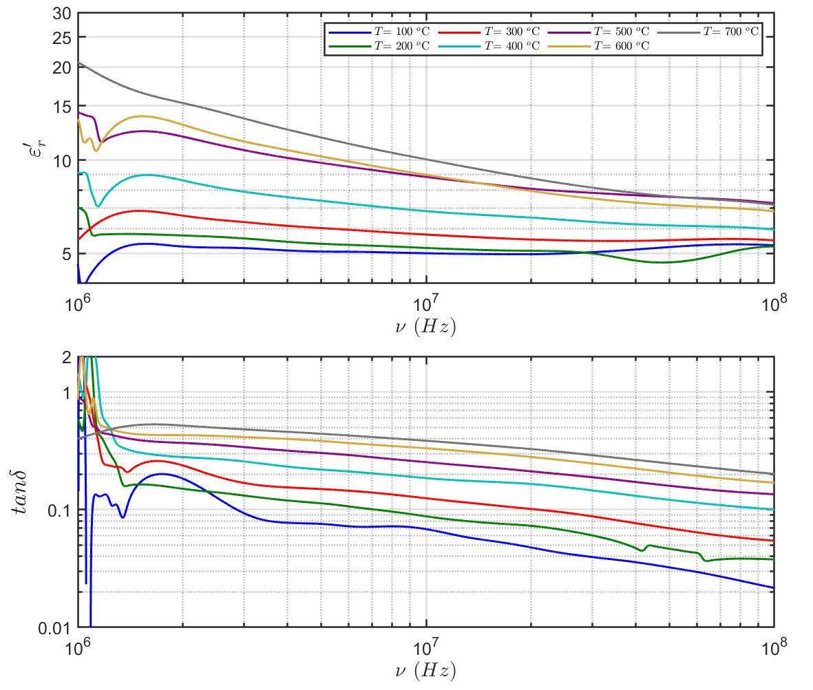
These measurements provide key constraints for electromagnetic wave propagation models and support the interpretation of future EnVision SRS observations. As an example, for a basaltic trachyandesite analogue, Figure 1 presents the real part of the complex relative permittivity (ε′ᵣ) and the loss tangent (tan δ) as functions of frequency at various temperatures. The results demonstrate a strong temperature dependence of dielectric properties: both ε′ᵣ and tan δ increase significantly with temperature, particularly at low frequencies, with no evidence of a relaxation mechanism across the measured frequency range.
The complex dielectric permittivity is defined as
The temperature dependence following an Arrhenius-type relation can be formulated as (Parkhomenko, 1967; Jonscher, 1981)
where and are coefficients, here E is activation energy in eV, eV/K is Boltzmann's constant, and T is absolute temperature in Kelvin. This formulation enables modeling of dielectric properties under Venusian thermal conditions.
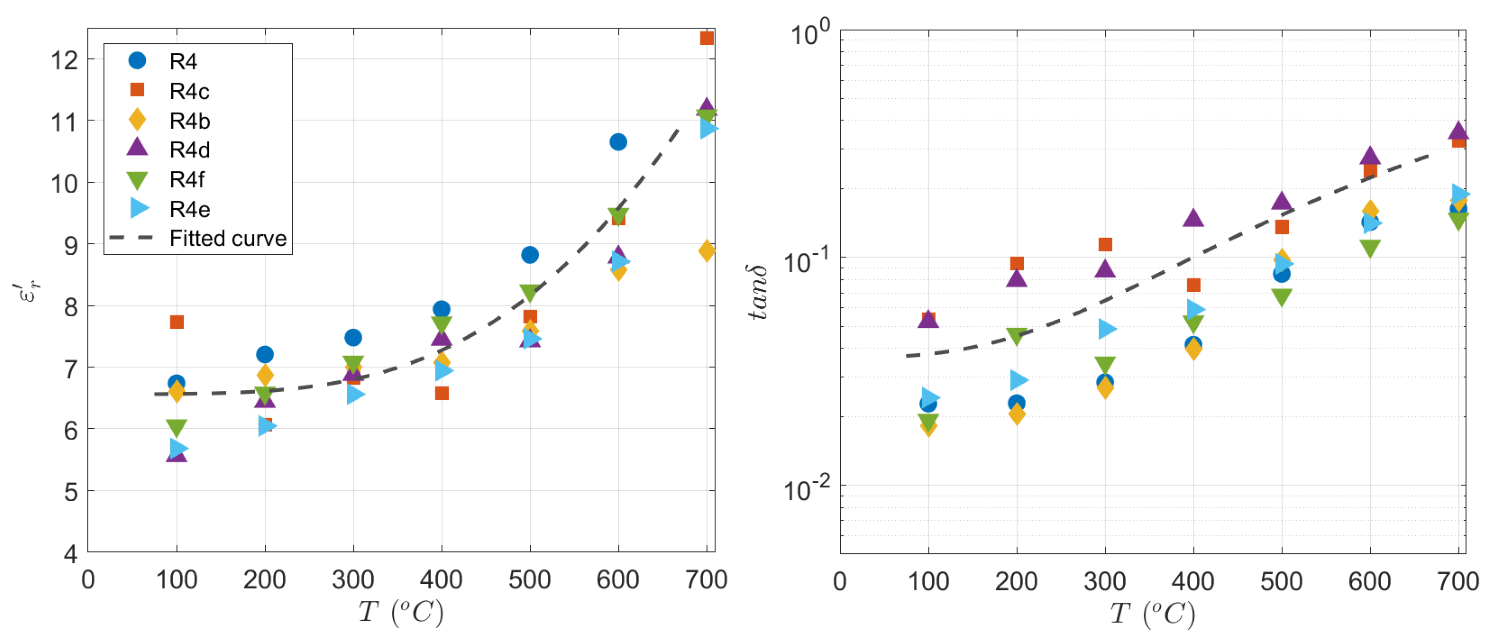
Figure 2 shows the dielectric properties of basaltic trachyandesite and rhyolite analogues at the SRS operating frequency (9 MHz). The loss tangent increases by up to an order of magnitude across the investigated temperature range. This behavior follows an Arrhenius-type relationship, allowing estimation of activation energies for different lithologies. The results indicate a strong increase in attenuation with temperature, which reduces radar signal penetration depth under Venus surface conditions.

Figure 1. Dielectric properties of Venusian analogues as a function of temperature and frequency: (top) real permittivity and (bottom) loss tangent.

Figure 2. Dielectric properties of basaltic trachyandesite analogues as a function of temperature at 9 MHz: (left) real permittivity and (right) loss tangent.
Acknowledgements
This work was supported by the Italian Space Agency (ASI) under the agreements Accordo Attuativo ASI–UNITrento/DISI n. 2022-23-HH.0 and Addenda n. 2022-23-HH.1-2023 and n. 2022-23-HH.2-2025, for the project “Attività scientifiche per il radar sounder di EnVision Fase B2/C.
References
- Jamaledin Baniamerian, Sebastian E Lauro, Barbara Cosciotti, Alessandro Brin, Carlo Lefevre, Elisabetta Mattei, and Elena Pettinelli. A new experimental setup for high-temperature dielectric characterization of Venus analogs. Journal of Geophysical Research: Planets, 130(7): e2024JE008545, 2025.
- I. Parkhomenko. Electrical Properties of Rocks. Springer, 1967. doi: 10.1007/978-1-4615-8609-8.
- K. Jonscher. The ’universal’ dielectric response. Nature, 267:673–679, 1977. doi: 10.1038/267673a0.
- Yu A Surkov, VL Barsukov, LP Moskalyeva, VP Kharyukova, and AL Kemurdzhian. New data on the composition, structure, and properties of venus rock obtained by venera 13 and venera 14. Journal of Geophysical Research: Solid Earth, 89(S02):B393–B402, 1984.
How to cite: Baniamerian, J., Emanuel Lauro, S., Rabiee, A., Cosciotti, B., Pettinelli, E., Marinangeli, L., Baliva, A., and Mattei, E.: High-Temperature Dielectric Characterization of Venusian Crust Analogues for Subsurface Radar Sounding, Europlanet Science Congress 2026, The Hague, The Netherlands, 7–11 Sep 2026, EPSC2026-69, https://doi.org/10.5194/epsc2026-69, 2026.
Please decide on your access
Please use the buttons below to download the supplementary material or to visit the external website where the presentation is linked. Regarding the external link, please note that Copernicus Meetings cannot accept any liability for the content and the website you will visit.
Forward to presentation link
You are going to open an external link to the presentation as indicated by the authors. Copernicus Meetings cannot accept any liability for the content and the website you will visit.
We are sorry, but presentations are only available for conference attendees. Please register for the conference first. Thank you.
Introduction: The Chang’E-5 lunar mission collected samples in the north-eastern region of Oceanus Procellarum in December 2020 (Li et al., 2022; Zhou et al.,2022). Two lunar samples from Chang’E5 mission (hereafter CE5-FR) were donated to France. These samples are curated at the Muséum National d’Histoire Naturelle (MNHN) in Paris to conduct non-destructive characterization. These analyses will provide reference data before their allocation to the French scientific community under the supervision of CNES. We will describe the general workflow and results obtained by the analysis of the container gas composition and by a non-invasive and non-destructive characterisation of individual particles extracted from both samples.
Samples and methods: The CE5-FR samples arrived in France stored in glass vials within two aluminium containers. The first sample was ~1 gram of surface-scooped sample (CE5C0100) and the second ~0.5 gram of drilled sample (CE5Z0800). Upon reception the samples were stored under ultra-dry and ultra-pure nitrogen (<1 ppm O2, <1 ppm H2O). X-ray Computed Tomography (XCT) allowed to perform 3D images of containers' inside components and to identify the samples' general features, which include tens of large grains (> 100 µm) and grains with higher density and heterogeneities (probably agglutinates).
Gas analysis: We developed a dedicated gas extraction apparatus to recover the containers' gas in the CE5-FR dedicated glovebox. The containers’ base was punctured and the extracted gas transferred into bottles cleaned under high vacuum. The operation was repeated for both CE5-FR drilled and scooped containers. The concentrations of H2O, O2, CO2, CO, and CH4 were quantitatively determined at LIPhy laboratory using a custom-built direct absorption laser spectrometer (H2O) and two ultra-sensitive cavity ring-down spectrometers (O2, CO2, CO, CH4) (Chaillot 2023). The argon concentration and isotopic compositions were measured at IPGP on a Noblesse HR 3F6M noble gas mass spectrometer. The results indicate that large amounts of O2 (~20.2%, ~13.9 %), CO2 (~840 ppm, ~640 ppm) and argon (>0.7%, >0.01%) were present in the containers for scooped and drilled sample, respectively, with values suggesting significant abundance of terrestrial atmospheric gases.
Bulk sample and individual particles analysis: The global sample mass was measured in the glove box using a Sartorius TE64 balance, and the magnetic susceptibility was measured, in collaboration with the CEREGE team, with a SM150L instrument in a magnetic field of 80 A/m and a variable frequency from from 63 Hz to 16 kHz.
During the gas extraction procedure, a small fraction of the grains escaped from the vials and was recovered on the surface of the Teflon support within each container. We extracted hundreds of grains in a cleanroom environment at IJCLab, and performed characterisation of these particles using complementary non-invasive techniques. Grains with sizes ranging from ~50 to 300 µm were individually documented using a Keyence VHX-7000 digital optical microscope, revealing a large variety of colors, transparency (from opaque to transparent), and morphologies, including smooth crystals, rough surfaces, inclusions, and spherical beads.
Fourier Transform micro-Infrared characterisation (FTIR) of these individual particles was performed with the I2SES bench at IAS. FTIR spectra were measured in reflection mode with respect to gold standards, using a Bruker LUMOS II micro-spectrometer in the mid-infrared: 2.5- 15 µm (4,000-670 cm-1) with a typical 50 x 50 µm spot size and spectral resolution of 4 cm-1. A subset of particles was analysed using a Renishaw InVia Raman microspectrometer using a 532 nm laser. High-resolution XRD was performed at IMPMC using a Bruker D8 diffractometer equipped with a double set of Mo/Ag anodes and focussing optics, a large C28 Photon 3 CMOS detector and a unique motorized goniometer allowing microXRD mapping experiment with a ~100µm beam size at the sample position.
A fraction of the scooped sample (~90 mg) and one millimetre-sized grain were extracted in the CE5-FR glovebox and analysed at IAS for microscopic hyperspectral near-infrared (NIR) spectroscopic analysis by PTAL/MicrOmega under controlled atmosphere (Loizeau et al. 2021). A similar analysis will be performed on the drilled sample and the detailed results will be presented in another contribution (see T. Jiang et al. this conference).
Some particles with smaller sizes were identified on the metallic support used for vial manipulation of drilled sample in the glove box. We recovered these particles with conductive carbon tape and they were carbon coated for analysis by scanning electron microscopy (SEM) and later transmission electron microscope (TEM) investigations. The SEM images were performed using SEM-TESCAN instrument at MNHN, allowing to obtain secondary electrons (SE) and back-scattering electrons (BSE) images together with semi-quantitative analysis of major elements by X-ray energy dispersive spectroscopy (EDS).
Further analysis and sample preparation will be performed using a micromanipulator in a dedicated cleanroom at IPGP. A sub-fraction of the sample will undergo gamma-ray spectrometry at the CEA/LNHB to produce additional data to aid the interpretation of radon measurements made by Chang’E-6/DORN (Chacartegui Rojo et al. 2025, Meslin et al. 2025).
The overall dataset of individual particles and bulk batches will be assembled in a database that will be made available under CNES supervision.
Acknowledgments:
We thank CNSA and the Chang’E-5 mission project for the gift of these lunar samples. This work was supported by DIM ACAV+ (Région Ile de France, C3E project), CNES (APR CE5-CURE), the MARCUS project (ANR-22-EXOR-0010, PEPR Origins). The I2SES bench was funded by DIM-Origins (I2SES project, PI. R. Brunetto), by MNHN, and ANR project LARCAS (grant ANR-22-CE49-0009). The work used the MNHN AST-RX platform for XCT analyses, TESCAN SEM microscope, and the MYRTHO cleanroom at IJCLab (supported by CNES). G.A was supported by the European Research Council (grant agreement n°101041122, project ATTRACTE). S.K. and H.F. thank the REFIMEVE network supported by ANR-11 EQPX-0039 and ANR-21-ESRE-0029.
References:
Li et al. 2022, National science review nwab188. Zhou et al. 2022, Advances in Space Research 823-836. Loizeau et al. 2020 PSS 193, 105087. Chacartegui Rojo et al. 2025, Applied Radiation and Isotopes 226, 112164. Meslin et al. 2025, EGU General Assembly, EGU25-14816. Chaillot, et al. 2025, Chemical Geology 673, 122450.
How to cite: Duprat, J. and Jiang, T. and the CE5-FR team: Curation of Chang’E-5 samples in France : containers opening and individual particle characterization, Europlanet Science Congress 2026, The Hague, The Netherlands, 7–11 Sep 2026, EPSC2026-770, https://doi.org/10.5194/epsc2026-770, 2026.
Introduction: China’s Chang’E-5 (CE-5) mission successfully returned ~1.73 kg of lunar samples from the northeastern region of Oceanus Procellarum in 2020 (Li et al., 2022; Zhou et al., 2022). These mare basalts (Morota et al., 2011; Qian et al., 2021; Che et al., 2021) originate from higher latitude than those explored by NASA’s Apollo and the USSR’s Luna missions, offering a unique opportunity to study previously unexamined lunar materials. As a state gift, France received 1.0 g of surface-scooped (CE5C0100) and 0.5 g of drilled material (CE5Z0800). Currently stored at the Muséum National d’Histoire Naturelle (MNHN) in Paris in a dedicated glovebox under ultra-pure nitrogen (<1 ppm O2 and H2O), a curation program was initiated by CNES to conduct non-invasive and non-destructive preliminary characterization of the samples before their allocation to the French scientific community.
Samples and methods: A ~90 mg powder (sub-bulk hereafter) and one millimeter-sized grain from the scooped sample were extracted into a sapphire dish within a Faculty-to-Faculty-Transfer-Container (FFTC) (Ito et al., 2020; Yada et al., 2023) inside the glovebox. Then the FFTCs were transported from MNHN to IJCLab for detailed optical imaging of the sample performed with a Keyence VHX-7000 in the MYRTHO cleanroom then transferred to the dedicated glovebox of the MicrOmega instrument installed on the PTAL setup at IAS (Bibring et al. 2017, Loizeau et al. 2021). The hyperspectral MicrOmega microscope covers the near-infrared range (0.99–3.65 µm,) with a spatial resolution of ~20 µm/pixel and a ~5 mm x 5mm field of view. Measurements were conducted in the glovebox under controlled pure-N2 (with 150-250 ppm H2O and 40-80 ppm O2).
Results: Optical imaging shows that the sub-bulk sample consists of fine gray powder (a few to a few tens micron in size) with occasional bright, larger particles. The average spectrum of the sub-bulk powder at the mm-scale exhibits a red slope typical of the lunar regolith (e.g., Pieters and Nobel 2016), starting from ~8% at 1 µm to ~18% at 2.5 µm. The spectrum also shows the olivine and pyroxene absorption feature (a right shoulder at 1 µm), and shallow absorptions around 1.3 µm, related to olivine and possibly plagioclases, and at 2 µm (pyroxene). These features are consistent with previous studies on other scooped sample from Chang’E-5 mission (e.g., Wu et al., 2024; Jiang et al., 2026). Notably, we observed a clear asymmetrical 3 µm band (centered at ~ 2.83 µm) related to H2O with a band depth is similar to that of Chang’E-5 scooped sample measured previously under air condition (Jiang et al., 2026). At the sub-mm scale, spectra of the sub-bulk powder reveal olivine (strong 1 µm absorption), high-Ca clinopyroxene (~2.2 µm absorption), and low-Ca orthopyroxene (~1.9 µm absorption) with size varying from a few tens µm to 200-300 µm. The 3 µm absorption related to H2O also shows some spatial variation. The large mm-sized grain reveals a complex mixture of mineralogical compositions. Detailed statistical analysis of these results will be presented at the conference.
Future plan: Ongoing work includes the analysis of the drilled sample, with similar measurements as for the scooped sample, on a fraction of the bulk powder and on mm-sized grains. We will compare the mineralogy and maturity of the samples between the two sampling sets. Additionally, we recovered small grains, with size between 50 and 300 µm, from the Teflon support during the opening of the containers. We characterized them with both the optical microscope and the FTIR I2SES facilities microscope at IAS (mid-infrared: 2.5-12.5 µm (4,000-800 cm-1) (Duprat et al. this meeting). The larger grains will be selected for MicrOmega measurements, which will allow to link the NIR and MIR data to provide better reference information for the grain composition.
Acknowledgements:
We thank CNSA and the Chang’E-5 mission project for the gift of these lunar samples, DIM ACAV+ (Région Ile de France, C3E project), CNES (APR CE5-CURE and MicrOmega development and activities), the MARCUS project (ANR-22-EXOR-0010, PEPR Origins) for financial support. The FTIR microscope at IAS was funded by DIM-Origins (I2SES project, PI. R. Brunetto). This work used the MYRTHO clean room at IJCLab, which receives support from CNES.
References:
Bibring, J-P., et al. 2017, Space Science Reviews 208, 401-412. Che, X., et al. 2021 Science 374, 887-890. Ito M. et al., 2020 Earth, Planets and Space 72:133. Jiang T. et al., 2026 A&A 705 A223. Li, C., et al. 2022, National science review nwab188. Li, Q., et al. 2021, Nature 600, 54-58. Loizeau et al. 2020 Planetary and Space Science 193, 105087. Morota, T., et al. 2011, Earth and Planetary Science Letters 302, 255-266. Qian, Y., et al. 2021, Earth and Planetary Science Letters 561, 116855. Wu, Y. Z., et al. 2024 A&A 682, A112. Yada T. et al., 2023 Earth, Planets and Space 75: 170. Zhou, C., et al. 2022, Advances in Space Research 69, 823-836.
How to cite: Jiang, T., Loizeau, D., Poulet, F., Pilorget, C., Brunetto, R., Duprat, J., Engrand, C., Dalauche, L., Bibring, J.-P., Hamm, V., and Lourit, L.: Near-Infrared Hyperspectral Imaging of Chang’E-5 samples in France using MicrOmega: Preliminary Results., Europlanet Science Congress 2026, The Hague, The Netherlands, 7–11 Sep 2026, EPSC2026-647, https://doi.org/10.5194/epsc2026-647, 2026.
Several approaches are currently available for investigating the composition of Mars and the Moon, including in situ analyses performed by rovers and landers, orbital observations, and laboratory investigations of meteorites collected on Earth. In the case of Mars, missions such as the Perseverance rover from the Mars 2020 mission [1] and the future Rosalind Franklin rover from the ExoMars mission [2] provide in situ geochemical and mineralogical analyses, while orbital studies are carried out by spacecraft such as the Mars Reconnaissance Orbiter [3]. Similarly, the Moon is being investigated through surface missions involving rovers such as the Yutu-2 [4], and the Pragyan rovers [5], together with orbital measurements acquired by missions including the Lunar Reconnaissance Orbiter [6]. Complementary information about these planetary bodies can also be obtained through the analysis of Martian and Lunar meteorites recovered on Earth. Their study also helps to better understand the limitations and constraints associated with different analytical techniques when applied to such complex and heterogeneous materials.
Meteorite samples are commonly prepared as polished subsamples suitable for non-destructive spectroscopic analyses. However, different preparation and preservation procedures can be applied, ranging from polished standalone pieces to subsamples embedded in resin blocks for improved mechanical stability and long-term preservation [7]. In this work, Martian and Lunar meteorites (LAR 12011, MIL 090036, MIL 05035 and EET 79001) prepared following these different methodologies were analyzed using micro-Raman spectroscopy, near-infrared (NIR) imaging, and micro-Energy Dispersive X-ray fluorescence (µED-XRF) in order to evaluate the influence of sample preparation on the measurements obtained with each analytical technique and to study and understand the limitations of these techniques.
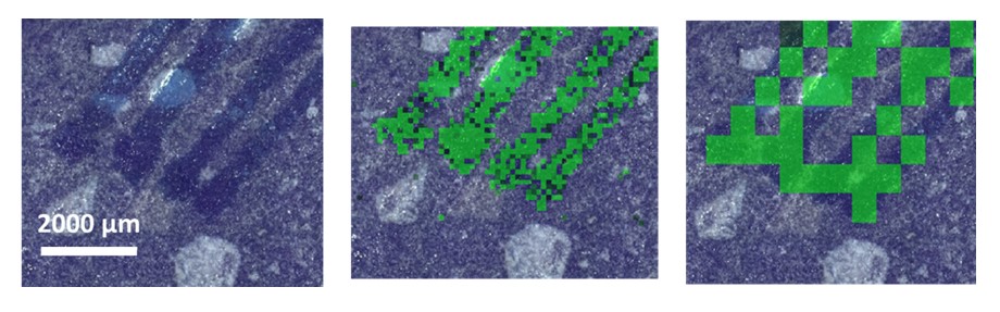
Particular attention was given to mapping modalities that allow the acquisition of hyperspectral data cubes. The role of spatial resolution, one of the most critical parameters in the spectroscopic characterization of heterogeneous planetary materials, was investigated. In fact, depending on the selected spatial resolution, the analyses may either provide information representative of the bulk matrix composition or enable the identification of minor mineral phases and mesostasis components present in lower abundances within the meteorites. To gain a clearer understanding of how spatial resolution affects each analytical technique additional experiments were conducted on a reference matrix of a terrestrial igneous analogue with chemical alterations with a similar composition than the studied meteorites. An example of Raman images obtained with different spatial resolution can be seen in Figure 1.

Figure 1. Raman images in the terrestrial analogue with different spatial resolution parameters. The central image was obtained using a step size of 105 µm, whereas the image on the right was acquired with a step size of 600 µm.
The results highlight the importance of optimizing the spatial resolution according to the analytical objective and of combining complementary spectroscopic techniques to achieve a representative and comprehensive characterization of the heterogeneous mineralogical and chemical composition of Martian and Lunar meteorites, as each method provides distinct information on the samples. Guidelines are also provided to help avoid misinterpretation of observed features and ensure consistency between measured signals and actual sample characteristics.
Acknowledgements
Meteorites were provided by the US Antarctic Meteorite Program, through the loan agreement between NASA’s JSC and the UPV/EHU. This work has been supported through the PAMMAT project “Alteration processes in Mars and Moon meteorites, and Terrestrial Analogues at different environments: Mars2020, Rosalind Franklin and Returned Samples from Mars and Moon” (Grant No. PID2022-142750OB-I00), funded by the Spanish Agency for Research MCIN/AEI/10.13039/501100011033/FEDER-UE, and the Strategic Project “Study of Alteration Processes in Terrestrial and Planetary Materials” (Grant No. UPV/EHU PES 21/88).
References
[1] T. Fouchet, J.-M. Reess, et al., The SuperCam infrared spectrometer for the Perseverance rover of the Mars 2020 mission, Icarus 373 (2021) 114773. https://doi.org/10.1016/j.icarus.2021.114773.
[2] J.L. Vago, F. Westall, et al., Habitability on Early Mars and the search for biosignatures with the ExoMars rover, Astrobiology 17 (2017) 6-7. https://doi.org/10.1089/ast.2016.1533.
[3] R.W. Zurek, S.E. Smrekar, An overview of the Mars Reconnaissance Orbiter (MRO) science mission, J. Gephys. Res. Planets 112 (2007). https://doi.org/10.1029/2006JE002701.
[4] C. Li, D. Liu, et al., Chang’E-4 initial spectroscopic identification of lunar far-side mantle-derived materials, Nature 569 (2019) 378-382. https://doi.org/10.1038/s41586-019-1189-0.
[5] S.V. Vadawale, N.P.S. Mithun, et al., Chandrayaan-3 APXS elemental abundance measurements at lunar high latitude, Nature 633 (2024) 327-331. https://doi.org/10.1038/s41586-024-07870-7.
[6] R. Vondrak, J. Keller, G. Chin, J. Garvin, Lunar Reconnaissance Orbiter (LRO): Observations for Lunar Exploration and Science, Space Sci. Rev. 150 (2010) 7-22. https://doi.org/10.1007/s11214-010-9631-5.
[7] R. Harrington, K. Righter, Polished sample preparation without epoxy, 50th Lunar and Planetary Science Conference (2019) 2132.
How to cite: Coloma, L., Gorla, G., Sánchez-Goyenaga, M., Alberquilla, F., Aramendia, J., Arana, G., and Madariaga, J. M.: Challenges in Achieving Representative Multispectroscopic Analyses of Martian and Lunar Meteorites: Effects of Sample Preparation, Heterogeneity, and Spatial and Spectral Resolution, Europlanet Science Congress 2026, The Hague, The Netherlands, 7–11 Sep 2026, EPSC2026-1121, https://doi.org/10.5194/epsc2026-1121, 2026.
Volcanic glasses are widespread on the Moon and represent an important component of both pyroclastic deposits and the lunar regolith. These amorphous materials originate from explosive volcanic eruptions as well as from impact-related processes, and therefore preserve valuable information on the evolution of the lunar crust and mantle. Despite their relevance for the interpretation of remote sensing observations, amorphous silicate materials remain significantly underrepresented in current spectral libraries, especially when compared to crystalline phases. In this framework, laboratory investigations on well-characterized analogue materials are essential for improving the identification and interpretation of glass-rich planetary surfaces. In this work, we present a combined Visible-Near Infrared (VNIR, 400-2500 nm) and Mid-Infrared (MIR, 3000-25000 nm) spectral characterization of synthetic silicate glasses representative of lunar mafic to ultramafic compositions.
Four lunar analogue compositions derived from Apollo samples were synthesized in laboratory: a high-Ti mare basalt (MB), a low-Ti basalt (LTB), a KREEP basalt (KB), and an Apollo 15 green glass analogue (GG). The glasses were produced from high-purity oxide mixtures and subsequently separated into four grain size fractions (<38 μm, 38-63 μm, 63-106 μm, and 106-150 μm) to evaluate the influence of particle size on spectral behaviour.
Reflectance spectra were acquired using FTIR (Fourier Transform Infrared) spectroscopy across the VNIR and MIR spectral ranges. Additional XRPD (X-Ray Powder Diffractometry) analyses confirmed the predominantly amorphous nature of the synthesized materials, although the high-Ti MB sample displayed traces of crystallization, likely related to Fe-Ti oxide nucleation during quenching. Spectral analyses focused on the behaviour of albedo, spectral slopes, VNIR absorption bands, and MIR diagnostic features such as the Christiansen Feature (CF), Reststrahlen bands peak position (RBpeak), and Transparency Feature (TF).
In the VNIR domain, all samples display weak and broad absorption features centered near ~1100 nm and ~1900 nm, attributed to Fe2+ electronic transitions in octahedral and tetrahedral coordination. Compared to crystalline materials, these absorptions appear toned down and poorly resolved, reflecting the amorphous structure of the studied glasses. Spectral slopes and albedo are strongly affected by grain size, with finer fractions generally showing higher reflectance and steeper VIS slopes. However, significant compositional effects are also observed. In particular, the MB sample shows an anomalous spectral behaviour, characterized by increasing albedo toward coarser fractions and flattened VNIR slopes. The influence of composition was investigated through the SAT parameter (SiO2+Al2O3+TiO2), the SCFM index, and iron content. Overall, grain size exerts the dominant control on VNIR spectral variability, partially masking compositional trends.
In the MIR region, spectral behaviour is dominated by vibrational processes within the silicate framework and provides clearer compositional constraints. The CF position systematically shifts toward shorter wavelengths with increasing grain size and exhibits a strong correlation with parameters related to silicate network polymerization, particularly SAT and SiO2 content. The finest grain size fractions (<38 μm) show the best agreement with trends previously reported in the literature for synthetic silicate glasses. In contrast, the RBpeak position displays a weaker and more scattered compositional dependence, although partial correlations with the SCFM parameter can be recognized. A weak TF is observed exclusively in the finest grain size fractions, confirming the strong grain size control on this spectral feature and the generally weak MIR spectral contrast of low-alkali silicate glasses.
In conclusion, the results demonstrate that MIR spectral parameters, particularly the Christiansen Feature, provide more robust compositional constraints than VNIR spectral properties, whereas VNIR data remain useful for identifying iron-rich glassy materials and evaluating grain size effects. The dataset presented here extends current spectral libraries toward primitive lunar mafic compositions and contributes to improving the interpretation of orbital and in situ lunar observations. This work contributes to improving spectral libraries of amorphous planetary materials and supports future investigations of glass-rich volcanic and impact deposits on the Moon and other rocky bodies.
Acknowledgements
We acknowledge the support of ASI under the ASI-UniPg agreement 2019-2-HH.0.
How to cite: Scognamiglio, G., Pisello, A., Fastelli, M., Tiraboschi, C., Zinzi, A., and Perugini, D.: Spectral Characterization of Synthetic Silicate Glasses Analogous to Lunar Basalts, Europlanet Science Congress 2026, The Hague, The Netherlands, 7–11 Sep 2026, EPSC2026-1094, https://doi.org/10.5194/epsc2026-1094, 2026.
The supercritical fluid state is reached for certain compounds at high pressure and temperature, above their critical point. Under these conditions, such fluids combine the properties of gases (high diffusivity, low viscosity) and liquids (high density, strong solvating power), making them particularly suitable for the extraction of organic molecules from complex matrices, such as extraterrestrial samples. For instance, meteorites, and especially carbonaceous chondrites, can contain up to 4% organic matter1,2. Organic compounds such as amino acids, nucleobases, sugars, and carboxylic acids, considered among the “building blocks of life”, have been reported in meteorite sample3–5. To better understand the role of meteorites in the emergence of life on Earth nearly 4 billion years ago, their molecular characterisation is therefore essential. However, organic matter, already present in low quantities in carbonaceous chondrites, is often strongly bound to the mineral matrix, making its extraction particularly challenging.
Supercritical fluids therefore offer a promising approach to overcome this limitation. The widespread use of supercritical CO₂ (scCO₂), due to its relatively low critical point (74 bar and 31 °C), as well as its availability, non-toxicity, non-flammability, and easy removal upon depressurisation, makes supercritical fluid extraction (SFE) a green chemistry technique6. In addition, scCO₂, commonly used for the extraction of non-polar compounds, can be mixed with polar co-solvents such as methanol, ethanol, dichloromethane, acetonitrile, or water to enable the simultaneous extraction of polar compounds. Moreover, recent instrumental developments allow the mixing of co-solvents with scCO₂ upstream of the sample, making it possible to generate co-solvent mixtures, gradients, and extraction sequences without intermediate sample handling, thereby reducing the risk of contamination. These advances over the past decades make SFE a promising alternative to more conventional extraction methods. It often provides comparable or higher recovery yields7–10, while overcoming common drawbacks such as high temperatures and pressures, multistep protocols, the use of toxic or polluting solvents, and contamination risks.
In the context of astrobiology, SFE is particularly well suited for complex and valuable samples, as it is a semi-destructive technique that preserves the mineral matrix after extraction of soluble compounds, while providing efficient extraction and a low risk of contamination. Although astrobiology emerged in the late 1950s, this technique has only begun to be explored in this field over the past three decades11–15, especially for sample return or meteoritic samples. In this study, we focus on meteoritic samples by investigating the capabilities of SFE considering recent developments, particularly the possibility of implementing a one-step protocol with successive co-solvent mixtures or gradients within a 2 mL sample cell, a more realistic volume given the limited availability of meteoritic material. To develop our protocol, we first optimise the large number of parameters using a meteoritic analogue. Using methanol as a co-solvent, an initial screening of the system was performed by focusing on the four most influential parameters (pressure, temperature, static extraction time, and methanol/scCO₂ ratio), allowing us to explore the parameter space with only 19 experiments using a full factorial design. Finally, considering that different molecules are extracted under different conditions, we aim to optimise the protocol using co-solvent gradients to develop a single extraction workflow capable of efficiently targeting multiple molecular classes through successive extraction steps. We will present the first results obtained from this optimisation and compare them with classical extraction methods.
- Botta, O. & Bada, J. L. Extraterrestrial Organic Compounds in Meteorites. Surveys in Geophysics 23, 411–467 (2002).
- Scott, E. R. D. & Krot, A. N. 1.07 - Chondrites and Their Components. in Treatise on Geochemistry (eds Holland, H. D. & Turekian, K. K.) 1–72 (Pergamon, Oxford, 2007). doi:10.1016/B0-08-043751-6/01145-2.
- Burton, A. S., Stern, J. C., Elsila, J. E., Glavin, D. P. & Et., A. Understanding prebiotic chemistry through the analysis of extraterrestrial amino acids and nucleobases in meteorites. Chemical Society Reviews https://doi.org/10.1039/C2CS35109A (2012) doi:10.1039/C2CS35109A.
- Alexander, C. M. O., Cody, G. D., De Gregorio, B. T., Nittler, L. R. & Stroud, R. M. The nature, origin and modification of insoluble organic matter in chondrites, the major source of Earth’s C and N. Geochemistry 77, 227–256 (2017).
- Martins, Z. Organic molecules in meteorites. Proceedings of the International Astronomical Union 11, 411–415 (2015).
- Ahmad, T., Masoodi, F. A., A. Rather, S., Wani, S. M. & Gull, A. Supercritical Fluid Extraction: A Review. JBCC 5, 114–122 (2019).
- Sun, L. & Lee, H. K. Optimization of microwave-assisted extraction and supercritical fluid extraction of carbamate pesticides in soil by experimental design methodology. Journal of Chromatography A 1014, 165–177 (2003).
- Sporring, S., Bøwadt, S., Svensmark, B. & Björklund, E. Comprehensive comparison of classic Soxhlet extraction with Soxtec extraction, ultrasonication extraction, supercritical fluid extraction, microwave assisted extraction and accelerated solvent extraction for the determination of polychlorinated biphenyls in soil. Journal of Chromatography A 1090, 1–9 (2005).
- Hawthorne, S. B., Grabanski, C. B., Martin, E. & Miller, D. J. Comparisons of Soxhlet extraction, pressurized liquid extraction, supercritical fluid extraction and subcritical water extraction for environmental solids: recovery, selectivity and effects on sample matrix. Journal of Chromatography A 892, 421–433 (2000).
- Spack, L., Alvarez, C., Martins, J. M. F. & Tarradellas, J. Comparison of supercritical fluid extraction (SFE), Soxhlet and shaking methods for pendimethalin extraction from soils: effect of soil properties and water content. Journal of Contaminant Hydrology 33, 171–185 (1998).
- McCaig, H. C. et al. Supercritical Carbon Dioxide Extraction of Coronene in the Presence of Perchlorate for In Situ Chemical Analysis of Martian Regolith. Astrobiology 16, 703–714 (2016).
- Menlyadiev, M., Henderson, B. L., Zhong, F., Lin, Y. & Kanik, I. Extraction of amino acids using supercritical carbon dioxide for in situ astrobiological applications. International Journal of Astrobiology 18, 102–111 (2019).
- Sephton, M. A., Pillinger, C. T. & Gilmour, I. Supercritical fluid extraction of the non-polar organic compounds in meteorites. Planetary and Space Science 49, 101–106 (2001).
- Abrahamsson, V., Henderson, B. L., Zhong, F., Lin, Y. & Kanik, I. Online supercritical fluid extraction and chromatography of biomarkers analysis in aqueous samples for in situ planetary applications. Anal Bioanal Chem 411, 8091–8101 (2019).
- Franco, C. & Hintze, P. Instrument for Solvent Extraction and Analysis (ISEE) of Organics from Regolith Simulant Using Supercritical Fluid Extraction and Chromatography. http://hdl.handle.net/2346/73007 (2017).
How to cite: Bouhier, B., Bourmancé, L., Stalport, F., Cottin, H., and Azémard, C.: Supercritical Fluid Extraction of organic molecules from extraterrestrial samples: A new one-step protocol proposed, Europlanet Science Congress 2026, The Hague, The Netherlands, 7–11 Sep 2026, EPSC2026-851, https://doi.org/10.5194/epsc2026-851, 2026.
Understanding the formation and activity of comets is key to constraining the conditions of the early solar system. As a comet approaches the Sun, its surface water-ice undergoes sintering, forming interparticle necks that significantly alter the ice’s physical properties, including its tensile strength. Gundlach et al. (2018) showed that sintering can proceed at temperatures as low as ~160 K. In this work, we systematically investigate how sintering temperature and duration affect the tensile strength of cometary ice analogues and examine the samples under a low-temperature scanning electron microscope (SEM).
Following the methodology of Kreuzig et al. (2024), we produce granular ice disks at 77 K in a newly designed sample holder. These disks are transferred into a liquid-nitrogen-cooled vacuum chamber, where they are heated from 77 K to a target temperature between 160 K and 200 K, held for variable time intervals on the order of hours, and then cooled back to 77 K to preserve the sintered microstructure. Tensile strength is measured in situ at 77 K using the Brazilian Disk Test (BDT), implemented on a custom apparatus adapted by the author from the CoPhyLab cone-penetration setup (Figure 1).
To resolve the microstructural evolution, sintered samples are examined under a low-temperature SEM, quantifying neck sizes as functions of sintering temperature and time (Figure 2). The resulting dataset will clarify the relationship between sintering parameters and tensile strength, thereby improving models of comet surface mechanics.
Figure 1. Photomontage of the planned Brazilian Disk Test (BDT) setup, analogue to the setup used by Kreuzig et al. (2024). A motor drives a stainless-steel blade, mounted to a precision load cell, downward onto the ice-disk sample seated on the sample table. The load cell continuously records the applied force and registers the peak load at sample failure. Blade displacement is tracked by a camera imaging a small reference tab against an LED backlight strip.
Figure 2. SEM picture of granular ice. The ice was kept at 77K at all times, no sintering is expected here and not visible.
How to cite: Quade, M., Kreuzig, C., Knoop, C., Timpe, M., and Blum, J.: Experimental results from CoPhyLab: Influence of Low-Temperature Sintering on the Tensile Strength of Cometary Ice Analogues determined by BDT, Europlanet Science Congress 2026, The Hague, The Netherlands, 7–11 Sep 2026, EPSC2026-450, https://doi.org/10.5194/epsc2026-450, 2026.
DNA is ubiquitous to all life on Earth and is thus considered an indicator for a universal ancestor; however, the origin of this ancestor is debated. Possible origins include endogenous production, for example in the young Earth’s oceans around hydrothermal vents, or exogenous delivery, as a result of impacts early in the Earth’s history [1]. If the former is correct, then Enceladus, with its briny liquid ocean, high geologic activity resulting in hydrothermal vents at the ocean-core boundary, and high abundance of complex organic molecules and fragments, is one of the key astrobiological targets within our Solar System [e.g., 2, 3]. However, its thick icy surface means it is not feasible for us to directly access and sample the ocean [4]; but conveniently, large plumes of ice and vapour are found to erupt from the Enceladean South Polar Region, potentially carrying a whole wealth of knowledge about the subsurface ocean [5]. As a universal feature of biological life on Earth, the detection of DNA in the ocean of Enceladus would provide strong indications of life within.
One of the biggest challenges for sample collection and analysis from the Enceladean plumes is capture speed, and thus peak impact shock pressure, which depend on the orbit type and is expected to range from 250 m/s (Enceladus orbit), up to 6 km/s or more (Saturn orbit/Enceladus flyby) [5]. Careful planning is therefore required to ensure efficient sample capture with minimal disruption or modification to minerals, organics, and biomarkers. Using the Light Gas Gun at the University of Kent, we are investigating the effect of impacts on the structure of DNA using a range of collection mediums, as well as different methods of post-impact recovery and analysis. At low speeds, up to ~ 1 km/s, short DNA ladders up to 1000 bp can survive and be characterised in analysis after impacting a soft metal such as indium. However, at higher speeds the concentration and structure of the DNA is lost. By contrast, DNA impacted onto aluminium is able to withstand speeds up to ~2.5 km/s, although there are potential signs of ladder breakdown. Further work at higher speeds is on-going to provide more definitive breakdown characterisation. Nevertheless, these preliminary results indicate that it would indeed be possible to successfully collect and characterise any small-chain DNA present in the plumes of Enceladus within current mission proposal constraints which include Enceladean orbiters.
[1] Chyba & Sagan, 1992, Nature; [2] Postberg et al., 2018, Nature; [3] Khawaja et al., 2025, Nature Astronomy; [4] Hemingway & Mittal, 2019, Icarus; [5] Burchell & Wozniakiewicz, 2024, MAPS.
How to cite: Grant, H., Robinson, G., Tandy, J., Burchell, M., and Wozniakiewicz, P.: Hypervelocity impacts of DNA on soft metals and implication for sample capture and characterisation efficiency , Europlanet Science Congress 2026, The Hague, The Netherlands, 7–11 Sep 2026, EPSC2026-1188, https://doi.org/10.5194/epsc2026-1188, 2026.
Please decide on your access
Please use the buttons below to download the supplementary material or to visit the external website where the presentation is linked. Regarding the external link, please note that Copernicus Meetings cannot accept any liability for the content and the website you will visit.
Forward to presentation link
You are going to open an external link to the presentation as indicated by the authors. Copernicus Meetings cannot accept any liability for the content and the website you will visit.
We are sorry, but presentations are only available for conference attendees. Please register for the conference first. Thank you.
Introduction: China's Tianwen-2 mission is designed to achieve orbital exploration and sample return from the near-Earth asteroid (2016 HO3), as well as a flyby exploration of the main-belt comet (311P), in a single launch [1]. Equipped with an Asteroid Thermal Emission Spectrometer (ATES), the mission aims to determine the surface mineral composition and thermophysical properties of both targets, thereby shedding light on their formation and evolutionary mechanisms [2]. However, on airless bodies like asteroids [3], the lack of interstitial gas limits heat transfer within the shallow subsurface to inefficient inter-particle radiation and contact conduction. This creates a steep thermal gradient within the top few hundred micrometers, which significantly alters thermal emission spectral features (e.g., shifting the Christiansen Feature (CF) to higher wavenumbers and increasing spectral contrast) [4-9]. Consequently, remote sensing thermal emission spectra cannot be directly interpreted using standard spectral libraries acquired under terrestrial conditions. To accurately interpret thermal infrared remote sensing data from airless bodies, laboratory thermal emission spectroscopy systems that simulate the environments of airless bodies are strictly required. Several such systems have been established internationally to simulate airless surface environments, including Brown University's ALEC [10], Oxford University's SLEC [11] and PASCALE [12], and Stony Brook University's PARSEC [13]. However, there is currently no comparable facility in China. To address this gap, this abstract introduces a newly, independently developed thermal emission spectroscopy measurement system (ChaSALE) designed for airless bodies such as the Moon and asteroids.
Design and Implementation of the ChaSALE: The ChaSALE primarily consists of a vacuum vessel, a 60K cryogenic helium circulation system, a sample cup assembly, a cryogenic off-axis parabolic mirror, a solar simulator and a control unit (Figure 1). The vacuum vessel provides the necessary vacuum environment for sample testing and includes a chamber, a pumping system and vacuum gauges. The system is capable of achieving a high vacuum of 8×10⁻⁹ bar inside the chamber. The 60K cryogenic helium circulation system cools the chamber to provide a cold background environment of < 100K, consisting of a thermal shroud, a cryocooler and associated piping. The sample cup assembly is used to hold and heat the test samples. From top to bottom, it comprises the sample receptacle, a heating module, a thermal insulation block, a support rod and a base. The heating module controls the sample temperature to simulate the varying thermal conditions on a planetary surface. The sample cup is designed with dimensions of Φ44 mm×5 mm. The cryogenic off-axis parabolic mirror collimates the divergent emission signals from the blackbody and the sample, directing the parallel beam into the spectrometer. A gold-coated aluminum mirror is utilized. To prevent the mirror's own thermal emission from contaminating the measurements, it is housed within a cold shield (Figure 1), which cools the mirror to approximately 150K during testing. The solar simulator is designed to replicate solar irradiation on the target body. The chamber is coupled to a Bruker Vertex 80V vacuum Fourier Transform Infrared (FTIR) spectrometer, which is equipped with a CsI beamsplitter and a liquid nitrogen-cooled Mercury Cadmium Telluride (MCT) detector. The operational capabilities and technical specifications of this system, alongside a comparison with similar international facilities, are summarized in Table 1.
Figure 1: Schematic diagram of the thermal emission spectrum measurement system and its optical path.
Table 1: Configurations and specifications of thermal emission measurement systems for the airless celestial bodies.
Samples Test: To validate the capability of our newly developed system to reproduce thermal emission spectra, we selected a particulate mixture of olivine and calcite (70 wt.% olivine and 30 wt.% calcite) with well-characterized spectral features as a test sample. We measured and derived the sample's emissivity under cryogenic vacuum conditions, and the results are presented in Figure 2. By comparing our data with standard mineral spectra for olivine and calcite, key diagnostic features can be clearly identified. For olivine, the CF at ~1100 cm-1, the Transparency Feature (TF) at ~790 cm-1, and the Reststrahlen Bands (RB) located between them are distinctly resolved. Similarly, for calcite, the prominent spectral features at ~875 cm-1 and ~710 cm-1, alongside the broad absorption feature between 1400 cm-1 and 1200 cm-1, are readily apparent. These findings successfully demonstrate the system's reliability in accurately reproducing the thermal emission spectra of geological samples under simulated cryogenic vacuum environments.
Figure 2: Validation results of the system’s ability to replicate sample thermal emission spectra.
References: [1] Zhang et al. 2021, Nature Astronomy, 5(8): 730-731. [2] Li et al. 2024, Journal of Deep Space Exploration, 11(3): 304-310. [3] Wang et al., 2019, Spacecraft Environment Engineering, 36(6): 533-541. [4] Logan et al. 1970, Journal of Geophysical Research, 75(32): 6539-6548. [5] Logan et al., 1973, Journal of Geophysical Research, 78(23): 4983-5003. [6] Henderson et al., 1994, Journal of Geophysical Research: Planets, 99(E9): 19063-19073. [7] Bishop and Moersch, 2020, Cambridge: Cambridge University Press. [8] Donaldson et al., 2017, Icarus, 283: 326-342. [9] Shirley and Glotch, 2019, Journal of Geophysical Research: Planets, 124(4): 970-988. [10] Bramble et al., 2019, Review of Scientific Instruments, 90(9): 093101. [11] Thomas et al., 2012, Review of Scientific Instruments, 83(12): 124502. [12] Donaldson et al., 2021, Journal of Geophysical Research (Planets), 126(2): e06624. [13] Shirley and Glotch, https:∥ui.adsabs.harvard.edu/abs/ 2015LPI....46.2025S/.
How to cite: Liu, B., Zhou, Q., Yang, S., Wen, W., and Li, C.: Design and Implementation of the Chamber for Simulating Asteroid and Lunar Environment(ChaSALE), Europlanet Science Congress 2026, The Hague, The Netherlands, 7–11 Sep 2026, EPSC2026-70, https://doi.org/10.5194/epsc2026-70, 2026.
Introduction: Visible and near-infrared (VNIR) spectroscopy is fundamental to our understanding of lunar surface mineralogy. Recent lunar missions, such as Chang’e-4 and Chang’e-5/6, have returned invaluable in situ rock spectral data and small rock chips. However, interpreting the spectra of intact rock fragments remains uniquely challenging. To supplement geographically limited returned sam-ples, lunar meteorites like Northwest Africa (NWA) 4734—a highly evolved mare basalt—serve as crit-ical natural laboratories. Extensive petrological studies indicate this meteorite has undergone se-vere impact metamorphism and multistage crystal differentiation[1-4]. Despite this wealth of geochem-ical data, laboratory VNIR spectral analyses of NWA 4734 rock samples remain scarce.
The Modified Gaussian Model (MGM) is usual-ly applied to the spectral interpretation of powder samples. By mathematically deconvolving complex, overlapping absorption bands into individual Gaussian distributions, the MGM allows us to es-timate the relative abundances of mafic minerals, such as the ratio of High-Calcium Pyroxene (HCP) to Low-Calcium Pyroxene (LCP)[5-7]. While the MGM has been successfully applied to planetary remote sensing[8-9], a major limitation exists: its calibration and validation have historically relied on crushed, well-sorted powder mixtures.
Applying the MGM to intact whole rocks—which feature complex multiple scattering and shock-altered phases—has rarely been systemati-cally tested. This study aims to bridge that gap. By utilizing high-resolution petrographic analyses to establish a definitive "ground truth" HCP propor-tion via image pixel counting, the MGM’s accuracy is directly evaluated when applied to the actual VNIR spectra of the NWA 4734 whole-rock slice, providing a vital baseline for future lunar rock spectral interpretations.
Samples and Analytical Methods: The lunar meteorite Northwest Africa (NWA) 4734, a basal-tic rock slice (~5 × 3 × 0.8 cm), was selected for this study due to its well-documented petrology and mineralogy. To establish ground truth for spec-tral interpretation, the same sample area was ana-lyzed using both image-based and spectroscopic methods. First, backscattered electron (BSE) imag-ing and energy-dispersive X-ray spectroscopy (EDS) were performed with a scanning electron microscope (SEM) to map mineral phases and identify high- and low-calcium pyroxenes (HCP and LCP)[3-4]. The proportion of HCP relative to total pyroxene was derived via pixel counting of BSE images after grayscale thresholding, with a 5% tolerance. Second, visible to near-infrared (VNIR) reflectance spectra (450–2500 nm) of the identical area were acquired using an ASD Field-Spec 4 spectrometer under darkroom conditions. The spectra were then decomposed using the Modi-fied Gaussian Model (MGM) to extract absorption band parameters (center, width, strength) for py-roxene and olivine. The ratio of band strengths at ~1 μm and ~2 μm was used to estimate HCP abun-dance[6]. This approach allows direct comparison between petrographically determined mineralogy and spectrally derived compositions.
Results and Discussion: 1) Petrographic Characteristics
BSE imaging reveals that NWA 4734 has a di-abasic texture, with plagioclase laths (5–10 μm) and larger, fragmented pyroxene grains filling tri-angular interstices. Almost all plagioclase is trans-formed into maskelynite, indicating severe impact metamorphism. Pyroxene grains exhibit two sets of near-orthogonal cleavages and impact melt pockets, while olivine shows a “Swiss cheese” texture. These features confirm that the meteorite experi-enced both intense shock and multistage magmatic differentiation, consistent with Apollo mare bas-alts[1].
2) Mineralogical Characteristics
EPMA data show that plagioclase is uniformly anorthitic (An84-91), while pyroxene compositions vary widely (Wo11-35En3-55Fs26-81), spanning augite and pigeonite. Olivine Fa contents range from 57 to 90. Both pyroxene and olivine display Mg-rich cores and Fe-rich rims, providing clear evidence of fractional crystallization. These heterogeneities support a complex magmatic history and are con-sistent with previous studies on NWA 4734[1,3].
3) Proportion of HCP from Image Pixel Counting
Pixel counting of 21 BSE images from the cir-cular area (later measured spectrally) yielded 1,913,565 LCP pixels and 3,635,247 HCP pixels, giving a raw HCP proportion of 65.5% (Figure 1). After converting 2D areal abundance to 3D volu-metric abundance using the power 3/2 transfor-mation[10], the HCP proportion becomes 72% ± 5.4%. This value serves as the petrographic ground truth for evaluating MGM performance on rock samples.
4) Proportion of HCP from MGM Deconvolu-tion
ASD reflectance spectra of the same area show absorptions near 1000 nm and 2200 nm, with asymmetry at 1000 nm. MGM fitting with eight Gaussians achieved RMSE < 1.4% (Figure 2). The band strength ratio (CBSR) at 1 μm between two pyroxene-related Gaussians is 0.869, correspond-ing to 67–75% HCP. The CBSR at 2 μm is 0.895, yielding 66–73% HCP (Figure 3). The average HCP proportion from MGM is 71% ± 10.1%, agreeing well with the pixel counting result.
5) Error Analysis and Applicability of MGM
Errors in pixel counting include a 5% tolerance in grayscale recognition and ~2% uncounted area (cracks, unresolved minerals), resulting in RMSE of 5.4%. MGM errors include spectral fitting (<1.4%) and the CBSR-HCP calibration (~10%), giving RMSE < 10.1%. Additional measurement mismatch between ASD and SEM areas contributes ~3% uncertainty. Within errors, the two methods yield comparable HCP proportions (72% vs. 71%), demonstrating that MGM is applicable to rock samples.
References: [1] Chen J. et al. 2019, JGRE, 124, 2583. [2] Elardo, S. M. et al. 2014, M&PS, 49, 261. [3] Wang, Y. et al. 2012a, M&PSA, 75, 5170. [4] Wang, Y. et al. 2012b, GeCoA, 92, 329. [5] Sunshine, J. M. et al., 1990, JGRB, 95, 6955. [6] Sunshine, J. M., & Pieters, C. M. 1993, JGRE, 98, 9075. [7] Sunshine, J. M., & Pieters, C. M. 1998, JGRE, 103, 13675. [8] Kanner, L. C., Mustard, J. F., & Gendrin, A. 2007, Icar, 187, 442. [9] Mustard, J. F., & Sunshine, J. M. 1995, Sci, 267, 1623. [10] Pieters, C. M. et al. 1993, JGRE, 98, 17127.
Figure 1: Calculating the proportion of HCP using the image pixel counting method. The pixels with the red colors represent HCP, and the pixels with the blue
colors represents LCP.
Figure 2: Spectral fitting results of NWA 4734 using MGM.
Figure 3: The logarithmical relationship between the CBSR and the proportion of HCP. (a) the relationship at the 1 μm region, (b) the relationship at the 2 μm region (Sunshine & Pieters 1993).
How to cite: Wen, W., Gao, F., Liu, B., Zhou, Q., Yang, S., and Li, C.: Applicability and accuracy assessment of the Modified Gaussian Model (MGM) on the rock samples’ spectral interpretation, Europlanet Science Congress 2026, The Hague, The Netherlands, 7–11 Sep 2026, EPSC2026-314, https://doi.org/10.5194/epsc2026-314, 2026.
Atom probe tomography (APT) is a powerful tool for the characterisation of materials at atomic scales. APT works by applying a pulsed voltage or pulsed laser to a needle like specimen that is 100 nm in diameter [1-2]. The voltage or laser pulse provides sufficient energy to field evaporate a single ion from the sample that then impinges on a position sensitive detector [1-2]. The time of flight provides elemental and isotopic information of the ion while the position of detection allows reconstruction of the ions original position in the sample generating a 3D atomic scale render of the material. Thus, APT provides unique information of a material’s structure and composition that is unobtainable in any other way [1-2].
Over the last decade APT has provided unique insights across a range of technological [1] and geological materials [2]. Applications in planetary science range from the detection of pre-solar nano-diamonds [3], to characterizing the nanoscale damage induced by space weathering [4-5], to trace element mobilization during asteroid impacts [6], to nanophase magnetic carrier phases in iron meteorites [7], and aqueous alteration of asteroids [8], among others. Recent successful asteroid sample return missions (JAXA’s Hayabusa and Hayabusa2 missions and NASA’s OSIRIS-REx mission) as well as planned future sample return missions to Mars, Phobos and the Moon mean that it is important to extract the most science from the minimum sample volume. APT represents the ideal tool in this field to maximize the scientific return while consuming very little (0.01 µm3) of these precious materials [9-10].
Excitingly, APT technology has greatly improved over the last few years with the development of deep-UV laser systems and cryo-vacuum transfer capabilities that will enable the characterization of traditionally challenging materials including many extraterrestrial materials such as delicate hydrous phases which is particularly important for characterizing water-rich C-complex asteroids such as Ryugu and Bennu as well as Mars Sample Return materials [9-10], as well as irradiated materials e.g., space weathered natural and anthropogenic materials. However, we have only scratched the surface of what is possible with the APT technique and many more applications are yet to be attempted [10]. With human ambitions reaching back to the Moon and the Artemis program aiming to establish a permanent human settlement it is vital to understand how space craft materials and components behave in the radiation-rich and harsh space environment beyond low Earth Orbit. APT is uniquely suited to provide an atomic eye view of these important processes.
We are excited to announce the installation of the Space Nanomaterials Atom Probe or SNAP laboratory at the University of Glasgow. SNAP will be the first APT facility in Scotland and the first worldwide to specialize in space science challenges. It will focus on providing atomic-scale structural details on materials for space applications including satellite structural materials, space-based batteries, microelectronics and radiation-hardened materials. The SNAP facility will comprise a CAMECA LEAP 6000XR system with combined voltage and deep UV laser pulsed operation capabilities, as well as a and cryo-vacuum transfer solution. These features make SNAP uniquely suitable for delicate samples and enabling, first of their kind measurements of challenging materials including space weathered satellites and extraterrestrial materials.
The SNAP facility is currently being commissioned and we anticipate ‘first-light’ in autumn 2026. We are already open to discussions for collaborative projects.
The SNAP facility is supported by dedicated research technical professional and expert research staff across a range of material science. As an international user-friendly facility, we also welcome collaborations from the international space science, materials science, planetary science and geological communities. Once the SNAP facility is operational, we will launch our access scheme to provide a collaborative route to use the latest APT technology. We look forward to working with you on this exciting new equipment, and please do not hesitate to reach out with any questions or research ideas.
References: [1] Gault B. et al., (2021) Nat. Rev. Met. Prim. 1(1) 51. [2] Reddy S.M. et al., (2020), G&GR, 44(1) 5-50. [3] Heck P.R. et al., (2014) MAPS, 49(3), 453. [4] Greer J., et al., (2020) MAPS, 55(2), 426-440. [5] Daly L. et al., (2021) Nat Astro. 5(12) 1275-1285, [6] Montalvo S.D., (2019) Chem. Geol., 507, 85-95. [7] Einsle J.F., et al., (2018) PNAS¸ 115(49) E11436-E11445. [8] White L.F., et a., (2020) PNAS, 117(21) 11217-11219. [9] Daly L. et al., (2020) IOP Conf. Ser. Mat. Sci. & Eng. 891(1), 012008. [10] McCarroll I.E. et al., (2022) MRS Bull., 47(7) 696-705.
How to cite: Daly, L., Maclaren, D., MacLaren, I., Bagot, P., Lee, M., Einsle, J., Ganin, A., and Mark, D.: The Space Nanomaterials Atom Probe (SNAP), Europlanet Science Congress 2026, The Hague, The Netherlands, 7–11 Sep 2026, EPSC2026-489, https://doi.org/10.5194/epsc2026-489, 2026.
Introduction
Martian analogue environments provide valuable opportunities to investigate the preservation of biosignatures and to refine strategies for their detection on Mars. Reactive gossans in permafrost - such as those found in the Canadian Arctic - constitute potential Martian analogues because of mineralogical similarities observed between terrestrial gossans and orbital or rover-based observations on Mars. Gossans are the superficial expression of sulfide-bearing rocks that have undergone chemical weathering through interaction with oxygenated groundwater. Gossans constitute one of the few iron-rich aqueous environments known to provide a habitable setting on Earth and potentially on Mars [1]. Previous studies have proposed that gossans, or related processes involving the chemical alteration of sulfide minerals, may have occurred on Mars in the past and that some of these reactions could persist today within the Martian permafrost [e.g. 2, 3, 4, 5].
The Terrestrial Mineral Analysis by Remote Sensing (T-MARS) project investigated Arctic gossans located at the head of Expedition Fiord, Axel Heiberg Island, Nunavut, Canada (Fig. 1) [6]. It included the detection and study of gossans from orbit to the ground using instrumentation relevant to present and upcoming Mars exploration missions. The scientific objectives were (1) to characterize their geochemistry and mineralogy; and (2) understand their formation processes. The operational objectives were (3) to develop strategies for detecting and studying them on Earth and Mars; and (4) to detect potential biosignatures.
Methods
Prior to field work
Before the field campaigns, target gossans were selected according to both their accessibility and their scientific relevance. A principal component analysis (PCA) was applied to a WorldView-2 image. A colour composite was then generated using the principal components showing the strongest contrast between the eigenvector values associated with the original red and blue bands referring to the red/blue ratio often used to detect iron oxides (Fig. 1). Previously identified gossan sites helped identify additional potential outcrops for field investigation on the colour composite. A slope map was used to assess the accessibility of the selected sites.
Field work
Two field campaigns to Expedition Fiord on Axel Heiberg Island, NU were carried out in July 2022 and July 2023 in the vicinity of the McGill Arctic Research Station. The investigated gossans were sampled both at the surface and at depth through vertical trenches. Both sterile and non-sterile sampling procedures were applied depending on the selected follow up laboratory protocols. A HySpex SWIR-384 hyperspectral camera was used to image gossan occurrences and evaluate the potential of this technology as a reconnaissance tool for mineral mapping. A drone was also deployed to examine hard‑to‑reach areas and to capture broader views of outcrops that were not visible from the ground (Fig. 2).
Laboratory analyses
Samples were characterized using elemental analyses for sulfur, nitrogen and carbon, together with X-ray diffraction and X-ray fluorescence techniques [7, 8, 9]. Additional measurements included loss on ignition, pH, and SEM-EDX analyses. Sterile samples were also investigated for lipid biosignatures through n-alkane quantification by gas chromatography–mass spectrometry [7], as well as for organic carbon content using Raman spectroscopy [8]. Spectroscopic measurements were acquired with rover-mountable instruments, including visible to near-infrared reflectance spectroscopy and mid- to thermal-infrared diffuse reflectance spectroscopy.
Remote sensing
The VNIR reflectance spectra were used as endmembers for the Spectral Feature Fitting algorithm in ENVI to identify gossan occurrences in WorldView-2 and PRISMA imagery [9].
Results and discussion
Elemental and spectroscopic analyses revealed mineral assemblages and oxidation gradients characteristic of gossans [10, 11, 12], with twenty-four mineral phases identified. Gypsum and quartz were common across all sites. Variations in primary sulfides and secondary sulfates indicate different oxidation stages, from actively weathering gossans to fully oxidized systems dominated by secondary sulfates. Organic carbon and potential lipid biosignatures were detected at depth. Classic gossan stratigraphy, with an oxidized surface layer over an unoxidized horizon, was observed at several sites, alongside additional stratification patterns ranging from distinct layering to strong heterogeneity (Fig. 3).
The results suggest that these gossans formed through alteration of primary sulfides in diabase/gabbro intrusions or from mobilized evaporitic sulfates, likely driven by paleohydrothermal activity or fluid circulation associated with the emplacement of sills in the host sedimentary rocks.
Field and laboratory observations made in this study indicate that successful identification of gossans on Mars will likely depend on targeting faulted regions where fracturing may have increased permeability, focusing on erosion-resistant ridges, using high-resolution orbital data and detecting co-located iron sulfides, oxides and sulfates.

Figure 1. Geology of the Expedition Fiord area, Nunavut. A) Location of Axel Heiberg Island in the Canadian Arctic Islands. B) Location of the McGill Arctic Research Station on Axel Heiberg Island. C) Footprint of the study area. Basemap for insets A-B-C: Esri World Imagery (2025). D) PCA colour composite. Figure modified after [6].

Figure 2: Multi-scale field images. A) Contextual overview of a sampling location acquired by drone. The white arrow indicates the gossan center shown in B (people present in the image for scale). B) Drone image of the gossan center, where a trench was excavated. C) Image captured by a team member on the ground showing gossan sampling. Figure from [6].

Figure 3: Stratification patterns observed in trenches. A) Classical gossan stratigraphy with unoxidized primary sulfide zone at depth and oxidized zone at the surface in a gossan (35 cm hammer). B) Finely stratified pattern of alternating oxidized and unoxidized layers in a gossan, measuring tape for scale (~60 cm deep). Figure from [6].
References
[1] Hays et al. (2017) Astrobiol., 17, 363-400.
[2] Burns, R.G. (1988) Lunar Planet. Sci. Conf. Proc., 18, 713–721.
[3] Dehouck et al. (2012). Geochem. Cosmochim. Acta, 90, 47–63.
[4] Moore and Szynkiewicz (2023) Icarus, 391, 115342.
[5] Gil-Lozano et al. (2025) Front. Astron. Space Sci. 12, 1504288.
[6] Lemelin et al. (2026) Planet. Space Sci., 106258.
[7] Aoid et al. (2023) Master’s degree thesis, McGill University.
[8] Belleau-Magnat et al. (2024) Planet. Space Sci., 256, 106036.
[9] Brassard et al. (2024) 55th LPSC, Abstract #1635.
[10] West et al. (2009) Planet. Space Sci. 57, 1302–1311.
[11] Peterson et al. (2014) Earth Planet Sci. Lett. 400, 88–93.
[12] Percival and Williamson (2016) Appl. Clay Sci. 119, 431–440.
How to cite: Belleau-Magnat, G., Lemelin, M., Williamson, M.-C., Léveillé, R., Brassard, É., Aoid, H., Fauconnier, B.-V., Douglas, P. M. J., Lachance, S., Marion, C. L., Castillo-Guimond, L., and Clark, S.: T-MARS Project: Reactive Gossans as Iron-Rich Martian Analogue Environments of Astrobiological Interest, Europlanet Science Congress 2026, The Hague, The Netherlands, 7–11 Sep 2026, EPSC2026-656, https://doi.org/10.5194/epsc2026-656, 2026.
The Lunar Regolith Simulant EAC-1 [1] is manufactured to imitate with high fidelity the composition and granularity of the Moon's surface. It is designed to be used in large amounts of several cubic meters, and already is in some locations.
Influence of the environment on the regolith is still possible, such as temperature and humidity variations of the diurnal or annual cycles. These variations, and the potential accumulation of residual humidity, i.e., humidity that infiltrates but cannot ever be removed, will over time change the electromagnetic properties of the regolith simulant. In order to ensure reproducible radar propagation measurements in a testing environment filled with regolith simulant, it is very useful to track the variation of the changes of the electromagnetic properties, based on easier available measurements of temperature and humidity. Also, the change of material's electromagnetic properties when an ice fraction is added to the mixture, is currently an open question.
Preliminary lab measurements show a dependency of the material's electromagnetic properties with respect to an applied mechanical compressive force. This observation is in line with measurements presented by other groups [2].
We will present permittivity measurements with systematically varied volume fractions of relative humidity and material density, in order to try to extract a numerical model for the material's behavior.
[1] V. S. Engelschiøn, et al., “EAC-1A: A novel large-volume lunar regolith simulant,” Sci Rep, vol. 10, no. 1, Art. no. 1, Mar. 2020, doi: 10.1038/s41598-020-62312-4.
[2] D. Ramos Somolinos et al., “Electromagnetic Characterization of EAC-1A and JSC-2A Lunar Regolith Simulants,” Materials, vol. 17, no. 15, p. 3633, July 2024, doi: 10.3390/ma17153633.
How to cite: Hegler, S., Benedix, W.-S., and Plettemeier, D.: Long-Term Environmental Influences on Lunar Regolith Simulant EAC-1, Europlanet Science Congress 2026, The Hague, The Netherlands, 7–11 Sep 2026, EPSC2026-754, https://doi.org/10.5194/epsc2026-754, 2026.
Europlanet provides access to state-of-the-art facilities for the simulation of planetary and wider space environments, analysis of planetary samples, testing and development of space technologies, and support of interdisciplinary studies.
Developed through European Commission funding from 2005-2024, and now operating as a self-sustaining non-profit association, Europlanet coordinates access to a global, distributed Research and Technology Infrastructure (RTI) that enables the planetary community to address major scientific and technological challenges. The facilities offered are owned and operated by organisational members of Europlanet, and cover a wide range of experimental set-ups and disciplines, ranging from neutron sources (which can be used e.g. for analysing crystalline and disordered structures in planetary/analogue samples) to light gas guns (to investigate hypervelocity impacts) and laboratories supporting techniques from electron-induced fluorescence (e.g. for studying the environments in planetary atmospheres) to carbon-14 dating accelerator mass spectrometry (e.g. for analyses of terrestrial/extraterrestrial organic and inorganic samples).
Europlanet’s flagship Transnational Access (TA) programme is designed to allow researchers from anywhere in the world, and at all career stages, to have access to facilities to carry out research projects and to foster international collaborations.
The TA programme supports all travel and local accommodation costs for European and international researchers to visit Europlanet’s RTI facilities for up to two weeks, as well as providing a contribution to the operating costs of the participating facilities. Proposals are evaluated by a peer review process.
The number and diversity of RTI facilities offered by Europlanet’s member organisations continues to grow rapidly. The 2026A call included 25 facilities hosted by ten organisations, and a second call with additional facilities is anticipated to open in September 2026.
Impact evaluation shows that access to facilities and expertise through Europlanet's TA programme leads to scientific advancements that would not otherwise occur, with over 90% of visits resulting in ongoing collaborations between the facilities and visitors and over 66% resulting in new avenues of research.1 Europlanet accelerates career development for early-stage researchers by providing training, mentoring, summer/winter schools, research project opportunities, and a professional network development.
In this presentation, we will give a status update on Europlanet’s distributed RTI and TA programme, as well as share plans for the future, including opportunities for access through future EC Grants.
- DeWitt, J., Heward, A. & Mason, N.J. Insights into evaluating a research project through an impact case study of a pan-European research infrastructure. Nature Astronomy , 9, 1415–1417 (2025). https://doi.org/10.1038/s41550-025-02684-7
How to cite: Heward, A., Mason, N., and Ivanovski, S.: Facility Access Through the Europlanet Distributed Research and Technology Infrastructure (RTI), Europlanet Science Congress 2026, The Hague, The Netherlands, 7–11 Sep 2026, EPSC2026-756, https://doi.org/10.5194/epsc2026-756, 2026.
Characterization of the geochemistry and organic content of Martian analog environments (reactive gossans) in the Canadian Arctic to assess their astrobiological potential.
1. Context
The past existence of potentially habitable environments on Mars [1-4] raises the question of whether any organic and mineralogical biosignatures have been preserved within the weathered Martian rock formations. However, the extreme physicochemical conditions prevailing on the surface of Mars, combined with oxidation and weathering processes, considerably complicate the identification and interpretation of such signatures [5-7].
In this context, the study of Mars analog environments is an essential approach for better understanding the interactions between geochemistry, mineralogy, and the preservation of organic matter under Mars-like conditions [5,8-11]. Among these, reactive gossans of the Canadian Arctic are of particular interest. These formations, resulting from the oxidation of sulfide deposits and subject to seasonal chemical weathering in a permafrost environment, constitute geochemically active environments capable of supporting sustained microbial activity [5,8,9,12-15]. Since similar conditions may have existed on Mars, these systems represent relevant analogs for studying Martian astrobiological potential and identifying preserved biosignatures.
This work aims to characterize the geochemistry and organic content of reactive gossans to assess their astrobiological potential and their relevance for interpreting future in situ observations of Mars.
2. Method
2.1 Field sampling
Two field campaigns were conducted on Axel Heiberg Island, Nunavut, to investigate six reactive gossans sites [12,13]. We focus on three of them: Color Ridge (CR-G3), White Glacier (WG-G1), and White Glacier Vein Array (WGVA-G1). Sampling was conducted at the surface and at depth to characterize the geochemical and organic evolution of the various strata. Six strata were sampled at CR-G3, four at WG-G1, and 12 at WGVA-G1. Sterile and non-sterile sampling protocols were applied depending on the planned laboratory analyses. Samples were stored at subzero temperatures to minimize the alteration of organic carbon.
2.2 Laboratory analyses
Quantitative and semi-quantitative characterizations included elemental analyses of sulfur, carbon, and nitrogen using an elemental analyzer; loss-on-ignition measurements to determine total organic carbon, total inorganic carbon, and total carbon; and pH
measurements in aqueous and saline solutions. Mineralogical and geochemical analyses were performed using WDXRF, XRD, and SEM-EDX. SEM-EDX analyses enabled the characterization of the elemental composition and mineralogy of structures or anomalies observed at the microscopic scale.
Qualitative characterizations were based on Raman spectroscopy, XRD, WDXRF, and SEM-EDX. Raman spectroscopy was used to identify mineral phases and to detect the D and G bands associated with the presence of organic carbonaceous material. An application was developed in-house to automate the interpretation of Raman spectra.
Some samples were metallized for observation under an SEM to identify any fossilized microstructures that might be associated with ancient microbial activity. This exploratory approach also aimed to assess the potential of this instrumentation for detecting morphological biosignatures in this type of Martian-analog environment.
3. Results
Preliminary results reveal significant geochemical, mineralogical, and organic heterogeneity among the three gossans studied. pH measurements show a marked contrast between the sites, with highly acidic conditions for CR-G3 (pH 2.2-2.7), acidic to near-neutral conditions for WG-G1, and neutral to slightly alkaline conditions for WGVA-G1. This variability reflects different degrees of oxidation and weathering within the profiles, linked to the reactivity of the sulfide phases.
Elemental analyses of sulfur, carbon, and nitrogen also reveal significant differences between sites and strata. CR-G3 exhibits the highest and most variable sulfur contents, suggesting the persistence of residual sulfides and/or the formation of secondary sulfates. Carbon contents measured by elemental analysis remain generally low, while nitrogen remains near detection limits, indicating a low contribution of nitrogenous organic matter. At the same time, loss-on-ignition measurements show mass losses that are sometimes more significant, which can be interpreted as a contribution from total organic carbon or carbonates depending on the temperature ranges considered. However, a discrepancy is observed between the results from elemental analysis and those obtained by loss on ignition. This difference indicates that the losses measured during heating do not exclusively reflect organic matter but may also be related to the dehydration of hydrated phases, the transformation of sulfates, or the decomposition of certain carbonate phases. These results highlight the need for a combined interpretation of elemental, mineralogical, and thermal data to better constrain the origin of mass losses and the actual preservation of organic carbon in these environments.
The WDXRF data indicate a composition dominated by SiO₂, Al₂O₃, and Fe₂O₃, with local enrichments in CaO, Na₂O, MgO, and SO₃ depending on the location. CR-G3 is characterized by high variability in Fe₂O₃, consistent with the oxidation of iron-rich sulfide phases. XRD analyses confirm this contrasting mineralogy, with a matrix dominated by
albite, with residual pyrite and secondary phases such as bassanite/gypsum, barite, and jarosite. The presence of jarosite is particularly significant, as it indicates acidic and oxidizing conditions relevant to altered Martian environments.
Raman and SEM-EDX analyses complement these observations by revealing fine mineral phases, Fe-S-rich structures, and potential carbon signatures, notably through the presence of D and G bands around 1350 and 1600 cm⁻¹, as well as light, carbon-rich structures observed in BSE. Filamentous or curved morphologies remain exploratory but constitute interesting targets for the evaluation of potential morphological biosignatures.
These results highlight the value of reactive gossans as Martian analogs for studying the relationships between sulfide weathering, pH gradients, the preservation of organic carbon, and the detection of potential biosignatures.
References
[1] Nazari-Sharabian et al. (2020) Galaxies, 8(2), 40.
[2] Martín-Torres et al. (2015) Nat. Geosci., 8(5), 357‑361.
[3] Ming et al (2007) https://ntrs.nasa.gov/citations/20070017324
[4] Palucis et al. (2014) J. Geophys. Res. Planets, 119( 4), 705‑728.
[5] Mustard et al. (2013) http://mepag.jpl.nasa.gov/reports/MEP/Mars_2020_SDT_Report_Final.pdf.
[6] Cockell et al. (2016) Astrobiology, 16(1), 89‑117.
[7] McMahon and Cosmidis (2022) J. Geol. Soc., 179(2).
[8] West et al. (2009) Planet. Space Sci., 57(11), 1302‑1311.
[9] Demaret et al. (2022) Astrobiology, 22(9), 1081‑1098.
[10] Fairén et al. (2010) Astrobiology, 10(8), 821‑843.
[11] Hays et al. (2022) Astrobiology, 17(4), 363‑400.
[12] Lemelin et al. (2026) Planet. Space Sci., 273‑274 (106258).
[13] Belleau-Magnat et al. (2025) Planet. Space Sci., 256(106036).
[14] Percival and Williamson (2016) Appl. Clay Sci., 119, 431‑440.
[15] Carman et al. (2024) ACS Earth Space Chem.,8(10), 1982‑1996.
How to cite: Fauconnier, B.-V., Lemelin, M., Charlier, B., Bonneau, A., and Belleau-Magnat, G.: Characterization of the geochemistry and organic content of Martian analog environments (reactive gossans) in the Canadian Arctic to assess their astrobiological potential., Europlanet Science Congress 2026, The Hague, The Netherlands, 7–11 Sep 2026, EPSC2026-777, https://doi.org/10.5194/epsc2026-777, 2026.
Introduction
Mid-Infrared (MIR) spectral analysis is one of the primary tools to studying the lunar soil mineral samples and providing a better understanding of their geological origin, helping to distinguish a mafic mare from a felsic anorthosite.
In this context, a set of 12 samples has been analyzed: one Apollo 16 sample (62231.44), endmembers (LHS-1 25A, LMS-1, LHS-1, LMS-1D, LHS1D) provided by the Exolith Lab of University of Central Florida, and intimate mixtures with different composition and same grain size (50%LMS1 + 50%LHS, 50%LMS-1D + 50%LHS1D), intimate mixture with same composition and different grain size (50%LMS1 + 50%LMS-1D, 50%LHS-1 + 50% LHS-1D), and intimate mixture with different composition and different grain size (50%LMS1 + 50% LHS-1D, 50% LHS-1 + 50% LMS-1D) under reflectance and in a vacuum environment at ambient temperature.
The primary goal is to characterize variations of spectral features such Christiansen Feature (CF) and Transparency Feature (TF) of these samples and see the relationship among them to understand how intimate mixing influences them. CF is sensitive to the presence of felsic minerals at shorter wavelengths and to the presence of mafic minerals at longer wavelengths, considering its region from 7.5 to 9.5 µm (Lucey et al., 2021). In our data, CF features are identified by the highest peak in this spectral window. The TF is an indicator of fine fraction samples where the mineral’s absorption coefficient reaches a minimum in the region between 11 and 13 µm (Prem et al., 2022).
Methodology
The set of samples has been analyzed in hemispherical reflectance spectroscopy under vacuum using a Bruker Vertex 80V FTIR spectrometer at the Planetary Spectroscopy Laboratory (PSL) at the German Aerospace Center (DLR). The set-up consists of a liquid-nitrogen-cooled MCT detector and KBr beam splitter operating under vacuum to simulate Lunar conditions. The measurements were taken under MIR range (approx. 7 to 25 µm). For each of the 12 samples, 3 measurements of 1000 scans were taken by rotating the sample approximately 120° between each measurement to provide a more accurate statistical analysis.
Pre-processing and data analysis
First, the reflectance data (R) from the spectrometer have been multiplied by a calibration factor of 0.93 to account for detector response. Then, the conversion from Reflectance (R) to Emissivity (E) has been applied as first approximation, according to Kirchhoff’s Law. For each sample, the three collected spectra were averaged. On each, the CF, TF and Reststrahlen Bands (RB) spectral features have been identified. A local polynomial fit (ranging from degree 4 to 12, depending on sample complexity) was applied to the CF window (7.5 - 9.5 µm) to get independence from random noise and maximize measurement precision. Furthermore, the Half Width Band Depth (HWBD) parameter, a physical parameter to quantify the volume scattering, has been identified for each of the TF feature of each sample to quantify the physical broadening of the absorption band, demonstrating the physical mechanism that “pushes” the CF towards a redshift. These results will focus on the quantification of the impact of fine particulates on mineralogical identification, possibly boosting further research and helping in the study of lunar soil, with a future perspective set on Moon expeditions.
References
Lucey, P. G., Greenhagen, B., Hanna, K. D., Bowles, N., Flom, A., & Paige, D. A. (2021). Christiansen feature map from the lunar reconnaissance orbiter diviner lunar radiometer experiment: Improved corrections and derived mineralogy. Journal of Geophysical Research: Planets, 126, e2020JE006777. https://doi.org/10.1029/2020JE006777
Prem, P., Greenhagen, B. T., Donaldson Hanna, K. L., Shirley, K. A., & Glotch, T. D. (2022). Modeling thermal emission under lunar surface environmental conditions. The Planetary Science Journal, 3(7), Article 180. https://doi.org/10.3847/PSJ/ac7ced
How to cite: D'Aniello, M., Barraud, O., Alemanno, G., Maturilli, A., and Donadio, C.: Mineralogical characterization of an Apollo 16 sample and lunar simulants through Mid-Infrared spectral analysis, Europlanet Science Congress 2026, The Hague, The Netherlands, 7–11 Sep 2026, EPSC2026-948, https://doi.org/10.5194/epsc2026-948, 2026.
Determining the diversity of exoplanets’ composition and structure is fundamental for investigating the Earth’s uniqueness. The improvement of ground-based instruments and space missions is and will provide unprecedented information as to exoplanet’s masses, radii and atmospheric speciation. However, none of these properties can uniquely constraint the nature of planetary interiors, which dictate their geological evolution. In order to interpret these new observations and identify future exoplanets targets, the implementation of accurate interior models is required. At present, much of our knowledge on exoplanets’ interior comes from analogue experiments in Earth-like composition performed over pressure-temperature-compositions spaces relevant for the Solar System. Stellar data have indicated a greater compositional diversity than in our Solar System. In this work we will present data from high pressure and temperature experiments conducted between 3 and 7 GPa to explore the potential mineralogical diversity of protoplanets. The bulk major elements composition was obtained from the variability observed in the host star catalogue, and the volatile content was changed in a systematic manner to cover the highest possible diversity. These experiments represent analogues for cores and mantles composition of planets orbiting stars with different compositions than the Sun and provide mineralogical data on potential diversity of exoplanets.
How to cite: Miozzi, F., Liebske, C., Sossi, P., and Alvaro, M.: Exploring the potential mineralogical diversity in bodies outside the solar system, Europlanet Science Congress 2026, The Hague, The Netherlands, 7–11 Sep 2026, EPSC2026-1110, https://doi.org/10.5194/epsc2026-1110, 2026.
With space agencies and private companies around the world expanding their activities in space, the Moon is now closer than ever before. NASA’s Artemis program missions are intended not only to return humans to the lunar surface, but also to establish a permanent presence there within the next decade. It is therefore essential to investigate the hazards present both in lunar orbit and on the surface in order to ensure astronaut safety. Impacts are among the most destructive processes in the Solar System and represent one of the major hazards on the lunar surface. Although catastrophic impacts, such as the asteroid event linked to the extinction of the dinosaurs, are extremely rare, the Earth–Moon system experiences a continuous flux of small meteoroids, ranging from decimetre-sized objects to dust particles.
Since the late 1990s, the lunar surface has been monitored to detect transient light phenomena produced during meteoroid hypervelocity impacts [1], commonly referred to as lunar impact flashes (LIFs). These observations have been conducted using small and medium-sized ground-based telescopes, with the aim of deriving the impact flux of centimetre- to decimetre-sized meteoroids [2–5]. In parallel, several detection and analysis techniques have been developed, depending on the observational setup (single- or dual-camera systems) and the scientific objectives of the observing teams [6,7]. Using data from NASA’s Lunar Reconnaissance Orbiter, several newly formed craters have been identified and linked to observed LIFs [2,8–11].
Observable impacts occur only every few hours, while more energetic events are even rarer and can only be detected under favourable observing conditions. Establishing a worldwide observing network is therefore essential to maximise monitoring time and improve detection statistics. The new Twin Impact Lunar Telescope (TILT) network will continuously monitor the Moon at visible and infrared wavelengths. The network will also support lunar seismology missions, including Chang'e 7, ESA’s LUMIO, NASA’s Farside Seismic Suite, and the South Pole Seismic Suite.
In preparation for these missions, and to support the interpretation of observations from LUMIO and the ground, we performed a series of laboratory impact experiments using the light-gas gun at the University of Kent to fire projectiles into lunar regolith simulants. The experiments were conducted over a range of impact velocities, while the resulting impact flashes were recorded using a custom-built spectrophotometric instrument with ten wavelength channels [10]. In this presentation, we will discuss our results.
Acknowledgments: This work is funded by STFC and UKSA grants. LUMIO is a mission funded under ESA’s General Support Technology Programme (GSTP) through the support of the national delegations of Italy (ASI), the United Kingdom (UKSA), Norway (NOSA), and Sweden (SNSA). The authors would like to acknowledge the support by the LUMIO Science Team.
References:
[1] J. L. Ortiz, P. V. Sada, L.R. Bellot Rubio, F. J. Aceituno, J. Aceituno, P. J. Gutierrez, U. Thiele, Optical detection of meteoroidal impacts on the Moon, Nature 405, 921–923 (2000).
[2] R. M. Suggs, D.E. Moser, W.J. Cooke, R.J. Suggs, The flux of kilogram-sized meteoroids from lunar impact monitoring, Icarus 238, 23–36 (2014).
[3] A. Z. Bonanos, C. Avdellidou, et al., NELIOTA: first temperature measurement of lunar impact flashes, A&A 612 (2018).
[4] C. Avdellidou & J. Vaubaillon, Temperatures of lunar impact flashes: mass and size distribution of small impactors hitting the Moon, MNRAS 484, Issue 4, p.5212-5222 (2019).
[5] C. Avdellidou, E. Munaibari, R. Larson, J. Vaubaillon, M. Delbo, P. Hayne, M. Wieczorek, D. Sheward, A. Cook, Impacts on the Moon: analysis methods and size distribution of impactors, Planetary & Space Science 200, 105020 (2021).
[6] E.M. Xylouris et al., NELIOTA: The wide-field, high-cadence, lunar monitoring system at the prime focus of the Kryoneri telescope, A&A, 619, id.A141, 14 pp. (2018).
[7] D. Sheward, M. Delbo, C. Avdellidou, A. Cook, P. Lognnone, E. Munaibari, L. Zanatta, A. Mercatali, S. Delbo, P. Tanga, Extending Lunar Impact Flash Observations into the Daytime with Short-Wave Infrared, MNRAS 529, Issue 4 (2024).
[8] D. Sheward, C. Avdellidou, A. Cook, E. Sefton-Nash, M. Delbo, B. Cantarella, L. Zanatta, PyNAPLE: Lunar Surface Impact Crater Detection, MNRAS 514, Issue 3, pp.4320-4328 (2022).
[9] D. Sheward, M. Delbo, C. Avdellidou, A. Cook, P. Lognonne Detection of small fresh craters on the Moon: Linking fresh craters to their lunar impact flash events, A&A, 699, id.L3, 7 pp. (2025).
[10] J. Tandy, M. C. Price, P. Wozniakiewicz, M. J. Cole, L. S. Alesbrook, C. Avdellidou, Impact flash evolution of CO2 ice, water ice and frozen martian and lunar regolith-simulant targets, MAPS 55, 10, 2301-2319 (2020).
How to cite: Avdellidou, C., Price, M., Sheward, D., and Tandy, J.: Laboratory investigation of lunar impact flashes in support for ground and space observations., Europlanet Science Congress 2026, The Hague, The Netherlands, 7–11 Sep 2026, EPSC2026-1113, https://doi.org/10.5194/epsc2026-1113, 2026.
The laser-induced liquid beam ion desorption (LILBID) experiment coupled with a time-of-flight (TOF) mass analyser, replicates mass spectra obtained from hypervelocity impacts of ice grains onto spaceborne instruments. LILBID has been used extensively to interpret mass spectra from Cassini’s Cosmic Dust Analyser (CDA) (Srama et al., 2004; Klenner et al., 2019), and has contributed to the characterisation of thousands of spectra collected during the mission. Thanks to the support of CNES, an Orbitrap-based laboratory testbench has been designed in collaboration with the Laboratoire de Physique et de Chimie de l’Environnement et de l’Espace (LPC2E).
The Orbitrap anaLYzer MultiPle IonizAtion (OLYMPIA) instrument has been successfully coupled to LILBID, achieving a resolving power of up to m/ΔmFWHM ~ 100000 at 19 u for a 500 ms FFT. This far exceeds the capabilities of time-of-flight mass spectrometers (m/ΔmFWHM ~ 600-800), thereby enabling the interpretation of spectral features without complications from isobaric interferences (Sanderink et al., 2023). This high resolution is essential for aiding the analysis of data from Europa Clipper’s SUrface Dust Analyzer (SUDA; Kempf et al., 2025).
In the near future, the coupling of OLYMPIA with LILBID will allow for the expansion of the mass spectral database of LILBID-TOF measurements (Klenner et al., 2022) in support of both Europa Clipper and ESA’s L4 mission to Enceladus. Thus far, OLYMPIA has been capable of detecting H3O+ and H2O+; however, prior to recent modifications, these measurements could only be linked to high-energy ionization exceeding 15 km/s (Sanderink et al., 2023).
A known marker for low-velocity impacts is the presence of water clusters in a gaussian distribution (Timmermann et al., 1991; Klenner et al., 2019). The new set-up, with capabilities for delayed extraction to simulate different ice grain impact speeds in space, aims to detect these water clusters while retaining the high resolution that characterizes Orbitrap mass spectrometry.
Various modifications and optimisation of the instrument have been performed at the LPC2E, aided by their experience with the LAb-CosmOrbitrap instrument, in support of the development of orbitrap-type instruments for space exploration (CosmOrbitrap) (Briois et al., 2016). In this context, the analytical performance of OLYMPIA has been assessed through UV laser ablation of an olivine sample (Figure 1). The achieved mass resolution for the calibrant peak, 24Mg+, reached m/ΔmFWHM ~ 225000 for 900 ms FFT (Figure 2). We present these new results, as well as the latest results for both mineral and icy analogue materials, which can also be useful in supporting the development of the CosmOrbitrap.
[Figure 1] UV laser ionization mass spectrum of an olivine mineral using the OLYMPIA instrument.
[Figure 2] 24Mg+peak’s resolution with 900 ms FFT duration.
Briois, C., Thissen, R., Thirkell, L., Aradj, K., Bouabdellah, A., Boukrara, A., ... & Makarov, A. (2016). Orbitrap mass analyser for in situ characterisation of planetary environments: Performance evaluation of a laboratory prototype. Planetary and Space Science, 131, 33–45. https://doi.org/10.1016/j.pss.2016.06.012
Kempf, S., Tucker, S., Altobelli, N., Briois, C., Cable, M. L., Grün, E., ... & Curtin, A. (2025). SUDA: A SUrface Dust Analyser for compositional mapping of the Galilean moon Europa. Space Science Reviews, 221(1), 10. https://doi.org/10.1007/s11214-025-01134-0
Klenner F, Postberg F, Hillier J, et al. Analogue spectra for impact ionization mass spectra of water ice grains obtained at different impact speeds in space. Rapid Commun Mass Spectrom. 2019;33:1751–1760. https://doi.org/10.1002/rcm.8518
Klenner, F., Postberg, F., Hillier, J., Khawaja, N., Reviol, R., Stolz, F., ... & Nölle, L. (2020). Analog experiments for the identification of trace biosignatures in ice grains from extraterrestrial ocean worlds. Astrobiology, 20(2), 179–189. https://doi.org/10.1089/ast.2019.2065
Klenner, F., Umair, M., Walter, S. H. G., Khawaja, N., Hillier, J., Nölle, L., ... & Postberg, F. (2022). Developing a laser induced liquid beam ion desorption spectral database as reference for spaceborne mass spectrometers. Earth and Space Science, 9(9), e2022EA002313. https://doi.org/10.1029/2022ea002313
Sanderink, A., Klenner, F., Zymak, I., Žabka, J., Postberg, F., Lebreton, J. P., ... & Briois, C. (2023). OLYMPIA-LILBID: A new laboratory setup to calibrate spaceborne hypervelocity ice grain detectors using high-resolution mass spectrometry. Analytical Chemistry, 95(7), 3621–3628. https://doi.org/10.1021/acs.analchem.2c04429
Srama, R., Ahrens, T. J., Altobelli, N., Auer, S., Bradley, J. G., Burton, M., ... & Zook, H. A. (2004). The Cassini Cosmic Dust Analyzer. Space Science Reviews, 114(1–4), 465–518.
Timmermann, R., & Grün, E. (1991). Plasma emission from high velocity impacts of microparticles onto water ice. In A. C. Levasseur-Regourd & H. Hasegawa (Eds.), Origin and Evolution of Interplanetary Dust (Vol. 175, pp. 375–378). Springer.
How to cite: Carmona Ruiz, M., Postberg, F., Briois, C., and Gaubicher, B.: Orbitrap-HRMS for calibration of hypervelocity ice grain space detectors: OLYMPIA, Europlanet Science Congress 2026, The Hague, The Netherlands, 7–11 Sep 2026, EPSC2026-1170, https://doi.org/10.5194/epsc2026-1170, 2026.
Introduction:
The most common type of rock present ont he surface of terrestrial planets in the Solar System are volcanic-magmatic rocks, which are constituted by lava flows and fragmented pyroclasts whose texture presents both glassy and crystalline silicate phases.
Planet Mars is, besides the Earth, the most diverse terrestrial planet in the Solar System, presenting magmatic rocks with compositions ranging from ultramafic/basaltic to alkaline/trachy-andesitic [1].
Understanding the influence that chemical composition and different phases (both crystalline and amorphous) have on the spectral response of volcanic rocks is pivotal to interpret remotely sensed spectra that are commonly used to interpret the geology of terrestrial planets. Thus, we synthesized Martian simulants with two putative Martian compositions, on which we performed cooling experiments together with a rheological, mineralogical and spectral characterization in order to provide reference to interpret the geological processes that have occurred on Mars.
Methods:
Samples were created by mixing powdered oxides to mimic the composition (Table 1) of volcanic products from Gusev and Gale craters, as they were hypothesized from data belonging to different missions [1].
Table 1: Composition of the starting materials accounted
|
|
Gusev |
Gale |
|
SiO2 |
47.33 |
52.04 |
|
TiO2 |
0.57 |
0.66 |
|
Al2O3 |
11.19 |
15.83 |
|
FeO |
19.12 |
10.89 |
|
MnO |
0.42 |
0.18 |
|
MgO |
10.29 |
4.44 |
|
CaO |
8.17 |
6.76 |
|
Na2O |
2.76 |
7.06 |
|
K2O |
0.14 |
2.14 |
The powders were molten at 1450°C to form a silicate melt, which were then quickly cooled to produce glasses. Then, for each of the produced glasses three cooling experiments were conducted in a Gero HTRV 70-250/18, equipped with Anton Paar rheometer [see 2 for reference]. Powdered glass was brought to superliqidus temperature and, after having reached the equilibrium, it was cooled down at a 100°C/h rate to crystallize. Crystallization was monitored through the measurement of viscosity during cooling, so that thre phases were individuated and samples: the point at which crystals start nucleating, the point at which crystal were grown and a final point of advanced crystallization where viscosity was not measurable anymore due to extreme rigidity of the samples (see arrows in Fig. 1).

Figure 1: Example of Viscosity path duting cooling for Gale simulant. Arrows indicate stages at which experiments were stopped.
The synthesized samples were analyzed using X-ray powder diffraction (XRPD), and quantitative phase analysis (QPA) was performed using the Rietveld profile fitting method with internal standards to determine amorphous content.
Spectral analyses:
Reflectance spectra were collected at the Cold Surface Spectroscopy (CSS) facility (https://cold-spectro.sshade.eu) located at the Institut de Pla-nétologie et d'Astrophysique (IPAG), Grenoble, France. The instrument used was the SHINE Spec-tro-Gonio Radiometer. The instrument is equipped with a cryogenic simulation chamber, CarboN-IR, to control the temperature of the samples. Spectra were collected in the 1-4.2 μm spectral range at different low temperatures between 105 and 290 K. The sample is cooled to a given temperature and then held for about 5–10 min to wait for thermal equilibrium and then during the measurement time, about 65–80 min depending on some acquisition parameters. We kept each sample for 70–90 min at a given temperature before starting our measurements.
Future perspectives:
Spectral characterization at low temperature is currently ungoing and will shed light on the influence of temperature on the detection of silicate phases. Preliminary spectral investigations suggest us that for samples with identical bulk chemical composition but different mineralogical assemblage, the spectral response within Visible and Near InfraRed (VNIR) and mid-infrared (MIR) is deeply different. VNIR spectra are influenced by the nucleation of iron-related phases whereas MIR spectra are dependant on Si-bearing phases. The mechanism influencing the shape of spectra results in fact from a complex interaction of the spectral response of different mineral/amorphous phases. In this way, the shape is easily misinterpreted as the features of the different phases which are often no longer recognizable. A thorough analysis helped us to understand which are the features that can be accounted for the interpretation of this kind of igneous materials, but a more comprehensive study on different compositions is needed for a complete assessment of this approach.
References:
[1] McSween Jr, H. Y. (2015). Petrology on mars. American Mineralogist, 100(11-12), 2380-2395.
[2] Vetere, F., Petrelli, M., Perugini, D., Haselbach, S., Morgavi, D., Pisello, A., ... & Holtz, F. (2021). Rheological evolution of eruptible Basaltic-Andesite Magmas under dynamic conditions: The importance of plagioclase growth rates. Journal of Volcanology and Geothermal Research, 420, 107411.
Additional Information: This work was carried on thanks to ASI-UniPG agreement 2019-2-HH.0 and in the framework of Trans-National Access research project selected and funded by Europlanet-2024 RI (European Union’s Horizon 2020 RI under grant agreement No. 871149). We also acknowledge the support from MUR in the framework of SUPER-C, ‘Dipartimento di Eccellenza 2023-2027’.
How to cite: Pisello, A., Maximiliano, F., Bernard, S., Pierre, B., Olivier, P., Francesco, V., Paola, C., and Diego, P.: Martian Simulants for Gusev and Gale craters igneous products: rheological, mineralogical and spectral characterization. , Europlanet Science Congress 2026, The Hague, The Netherlands, 7–11 Sep 2026, EPSC2026-1266, https://doi.org/10.5194/epsc2026-1266, 2026.
Please decide on your access
Please use the buttons below to download the supplementary material or to visit the external website where the presentation is linked. Regarding the external link, please note that Copernicus Meetings cannot accept any liability for the content and the website you will visit.
Forward to presentation link
You are going to open an external link to the presentation as indicated by the authors. Copernicus Meetings cannot accept any liability for the content and the website you will visit.
We are sorry, but presentations are only available for conference attendees. Please register for the conference first. Thank you.
You have already stored your personal programme. Please decide:
the present selections with my stored personal programmemy stored personal programme with the present selections
Please decide on your access
Please use the buttons below to download the supplementary material or to visit the external website where the presentation is linked. Regarding the external link, please note that Copernicus Meetings cannot accept any liability for the content and the website you will visit.
Forward to session asset
You are going to open an external link to the asset as indicated by the session. Copernicus Meetings cannot accept any liability for the content and the website you will visit.
We are sorry, but presentations are only available for conference attendees. Please register for the conference first. Thank you.